Structural Insights into the Peripheral Light-Harvesting Complex (LH2) in Purple Bacteria
50 likes | 183 Vues
The light-harvesting complex (LH2) is a nonamer exhibiting ninefold rotational symmetry, binding 27 Bacteriochlorophyll a (Bchl a) molecules and 9 carotenoids. Crystallographic studies reveal a 3-fold rotational axis, with symmetry preserved down to a resolution of 2.0 Å. The arrangement of pigments within the protein matrix is critical for the complex's optical properties, allowing the bacteria to efficiently harvest photons across various wavelengths. These findings enhance our understanding of the intricate structure-function relationship in photosynthetic organisms.

Structural Insights into the Peripheral Light-Harvesting Complex (LH2) in Purple Bacteria
E N D
Presentation Transcript
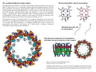
The peripheral light-harvesting complex The light harvesting complex is a nonamer with ninefold rotational symmetry. The molecule binds 27 Bchl a and we see 9 carotenoids (rhodopin glucoside) although estimates from the native membrane of the bacteria suggest that there ought to be 18. The crystal structure has a 3-fold rotational axis intersecting the molecule - so in the crystal 1/3 of the molecule is unique: the 9-fold molecular symmetry further reduces the structure to a unit of two polypeptide chains, 1 carotenoid and 3 Bchl a molecules. There are minor departures from this (non-crystallographic) 9-fold symmetry, notably at crystal contacts, but for the vast majority of the molecule this symmetry is precise down to our current resolution of 2.0 Å. The pigments are held by the protein matrix in a systematic arrangement. At the top of the molecule is an overlapping ring of Bchl a molecules. In the diagram below these are seen edge on in orange (only the chromophore, or conjugated double bond system, is shown). At approximately the middle of the structure are 9 further Bchl a molecules. Carotenoid chromophores span the gap between the Bchl a's. Bacteriochlorophyll a and its chromophore Rhodopin glucosideand its chromophore This physical arrangement of chromophores determines the gross properties of the complex http://www.chem.gla.ac.uk/protein/LH2/lh2struc.html Further information: steve@chem.gla.ac.uk Cogdell R.J., Isaacs N.W., Freer A.A., Arrelano J., Howard T.D., Papiz M.Z., Hawthorn-thwaite-Lawless A.M. & Prince S.M. (1997) The Structure and function of the LH2 (B800-850) complex from the purple bacteria Rhodopseudomonas acidophila strain 10050. Prog. Biophys. molec. Biol. Vol 68 No. 1. pp 1-27.
Carotenoid absorption (circa 470nm), Bchl a absorption at 800nm and Bchl a absorption at 850nm Energytransferin LH2 Photons may be absorbed by any of the pigments in the LH2 complex. Each pigment has characteristic resonant absorptions: Bchl a is a porphyrin like molecule with an asymmetric conjugated double bond system: this results in two resonant absorption bands. This asymmetry is the primary reason for the bacteria to choose this pigment. The organism needs to access photons from regions of the spectrum which have not been depleted by higher organisms - the asymmetry in the conjugated double bond system of Bchl a splits the resonant absorption bands of this molecule further than the (less asymmetric) Chlorophyll molecule. This allows the bacteria to find a niche in the spectrum in vivo. These absorptions may be mapped to transition dipoles (Qx and Qy) which can in turn be physically mapped onto the surface of the Bchl a molecule (the Q dipoles are approximately perpendicular and exist in the plane of the chlorin, Qy is coincident with the long axis of the chromophore).The carotenoid molecules in purple bacteria have a greater number of conjugated double bonds than those found in higher plants. This is a consequence of having to quench a triplet energy level in Bchl a for photoprotective purposes. However a consequence of the large extent of the chromophore is again access to a less depleted portion of the spectrum. In fact purple bacteria are purple due to the absorption of their carotenoids rather than Bchl a! Information from spectroscopy, biochemistry, and molecular biology has allowed us to assign bands in the LH2 spectrum to specific molecules in the LH2 molecule. A Qy absorption band occurring at 800nm is due to a monomeric Bchl a pigment oriented perpendicular to the membrane normal. A Qy absorption band occurring at 850nm may be assigned to extensively coupled Bchl a pigments with dipoles oriented parallel to the membrane normal. The Qx dipole absorptions of all Bchl a's are at the same wavelength.
Scanning Tunneling Microscopy, IBM Scientists discovered a new method for confining electrons to artificial structures at the nanometer lengthscale, the Quantum Corrals. Surface state electrons on Cu(111) were confined to closed structures (corrals) defined by barriers built from Fe adatoms. The barriers were assembled by individually positioning Fe adatoms using the tip of a low temperature scanning tunneling microscope (STM). A circular corral of radius 71.3 Angstrom was constructed in this way out of 48 Fe adatoms. This STM image shows the direct observation of standing-wave patterns in the local density of states of the Cu(111) surface. These spatial oscillations are quantum-mechanical interference patterns caused by scattering of the two-dimensional electron gas off the Fe adatoms and point defects.
for comparison rotate around 45° Progress of High Resolution Molecular Imaging and TEM Resolution Using 100 kV electron microscope (JEM-100B), molecular image of chlorinated copper phthalocyanine crystal was demonstrated that molecule could be observed by many beam imaging method for the first time in the world. After that, by increasing the acceleration voltage largely the resolution was improved in photographing the same sample with 500 kV High Resolution Electron Microscope (JEM-500, HAREM), which was presented at Novel symposium in 1979. The below figure was taken with new 1000 kV High Resolution Electron Spectromicroscope. The contrast inside the molecule becomes clearer, so that the benzen and the porphyrin rings appear clearer. Taken from http://eels.kuicr.kyoto-u.ac.jp