Download

1 / 34
340 likes | 379 Vues
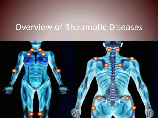
Learn to identify key features of rheumatology disorders at a glance! Recognize osteoarthritis, rheumatoid arthritis, psoriatic arthritis, Reiter's syndrome, lupus erythematosus, dermatomyositis, and more with visual cues and distinct symptoms.

E N D
High Impact RheumatologyRheumatology at a Glance Know It When You See It *
Know It When You See It Osteoarthritis: Typical hand • Hard boney enlargements • Heberden’s nodes at the DIP joints • Bouchard’s nodes at the PIP joints • Often have “squared” first CMC joint due to osteophytes at that joint
Know It When You See It • Rheumatoid arthritis • Soft synovial swelling • Synovitis and volar subluxation at the MCP joints • Synovitis of the wrists • Synovitis of the PIP joints with early swan neck deformities
Rheumatoid Arthritis: Swan Neck and Boutonnière Deformities • Late-stage findings indicating serious changes in the joints • Swan neck (digits 2 to 4) PIP extension DIP flexion • Boutonnière (digit 5) is the reverse; PIP flexion DIP extension
Know It When You See It • Tendon rupture in RA • Inability to extend fourth and fifth digits • Due to deformity and inflammation at the wrist causing excess wear of the extensor tendons
Know It When You See It • Psoriatic arthritis • Inflammation of the DIP joints • Sausage fingers • Joint involvement shows radial pattern • Nail changes • Psoriatic patches • Arthritis may start before the skin
Know It When You See It • Psoriatic arthritis • Sausage toes • IP joint involvement of a toe suggests a rheumatoid variant • Psoriatic arthritis and Reiter’s disease are the most common causes
Know It When You See It • Reiter’s syndrome • Keratoderma blennorrhagica • May look like psoriasis or syphilis • Can occur in patches or as sterile pustules
Reiter’s Syndrome (Reactive Arthritis) Seronegative asymmetric arthritis • Following: • Urethritis or cervicitis • Infectious diarrhea • Often associated with: • Inflammatory eye disease • Balanitis, oral ulceration, or keratoderma • Enthesopathy • Sacroiliitis
Inflammatory Bowel Disease • Ulcerative colitis • Regional enteritis (Crohn’s disease) • ? Whipple’s • ? Behçet’s
Know It When You See It Systemic lupus erythematosus • Butterfly rash • Involves cheeks and nose • Patient also has rash on chin and some telangiectasia
Know It When You See It Systemic lupus erythematosus • Interarticular dermatitis • Also has periungual erythema • This rash is distinct from that seen in dermatomyositis that occurs over the joints
Know It When You See It • Dermatomyositis • Scaly rash over the extensor surfaces of the interphalangeal joints
Know It When You See It • Periungual changes • Seen in lupus erythematosus, dermatomyositis, and scleroderma • Thickening of capillary loops • Dropout of capillary loops • Hemorrhage in the nail fold may also be present
Know It When You See It Close-up views of periungual changes Upper right: Dilated loops Upper left: Normal Lower right: Dilated loops with branching Lower left: Dilated loops with dropout View with ophthalmoscope and drop of oil
Know It When You See It • Dermatomyositis • Mantle or shawl distribution of rash
Know It When You See It • Linear scleroderma • Not usually associated with systemic disease
Know It When You See It • Livedo reticularis • Appears in a broad- based interrupted pattern in systemic vasculitis, including SLE • May occur as a fine, connected, lacy pattern in normals
Know It When You See It • Palpable purpura • Characteristic of dermal vasculitis in Henoch-SchÖnlein purpura
Know It When You See It • Saddle nose deformity • Relapsing polychondritis • May also occur in Wegener’s granulomatosis and syphilis
Know It When You See It Relapsing polychondritis Left: Ear changes with inflammation in the cartilage and swelling Right: Loss of ear cartilage in late stages
Know It When You See It • Ochronosis • Deposition of homogentisic acid • Gray discoloration of the ear and dense pigment on transillumination
Know It When You See It Gout tophi in the ear a good tip-off if present • Tophi appear rather late in gout • Prick the tophus with a needle. Put the drop of material on a slide • Multiple birefringent crystals will be seen on polarized microscopy
Know It When You See It • Urate crystal in a tophus • Top: Seen with ordinary light microscope with condenser racked down and light intensity adjusted • Bottom: Seen with compensated polarized light, the preferred method
Know It When You See It • Gouty tophus on finger • Note the yellow- orange color typical of a tophus • Patient also has swelling of the PIP of the index and fifth digits
Know It When You See It Skin pustule with disseminated gonorrhea • Usually a few lesions • Usually found on the extremities
Know It When You See It • Septic olecranon bursitis • Swelling of the bursa • Erythema and tenderness • If it looks ugly, tap it
Know It When You See It • Septic prepatellar • bursitis with cellulitis • Rubor, calor, dolor over the patella and adjacent tissue • Lack of joint involvement evident from nontender suprapatellar pouch and popliteal area • Don’t tap a normal knee through cellulitis
Know It When You See It Hypertrophic osteoarthropathy • Clubbing with loss of nail angle • Full syndrome includes periostitis of ends of long bones • Associated with • Chest malignancies • Chronic lung infection • Other tumors
Know It When You See It • Amyloidosis • Shoulder pad sign • The worst case you are likely to see • Patient also has macroglossia and purpura
Know It When You See It • Hyperthyroidism • Acropachy • Right: Soft tissue swelling between joints • Left: Periosteal new bone formation
Know It When You See It • Ehlers-Danlos syndrome • A true connective-tissue disease • Left: Hypermobility of joints. Can touch thumb to volar surface of forearm • Right: Hyperelasticity of skin • Associated with vascular abnormalities