Download
1 / 38
380 likes | 408 Vues
Ventricular resection tackles abnormal left ventricular contours to improve cardiac function. Learn about indications, surgical techniques, outcomes, and survival rates from key studies worldwide.

E N D
Ventricular resection in cardiac failure Surendra Naidoo Cardiothoracic Surgery Durban
Ventricular aneurysm resection Definition strictly - distinct area of abnormal left ventricular diastolic contour with systolic dyskinesia or paradoxical bulging loosely… intraoperative…
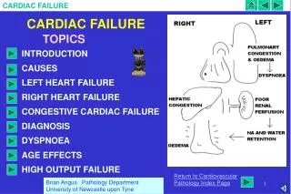
Pathophysiology Early expansion phase onset of MI gross thinning of infarct zone endocardium is smooth deposition of fibrin & thrombus Laplace's law (T = Pr/2h) Late remodelling phase 2 – 4 weeks after MI granulation tissue replaced by fibrous tissue mural thrombus
Indications for surgery Asymptomatic incidental during CABG moderate size CASS Study
Indications for surgery Symptomatic CHF angina arrhythmias embolism
TechniquesGeneral CPB VF cardioplegic arrest epicardial mapping Off-pump
Techniques Plication Linear closure Circular patch Endoventricular patch
Plication small aneurysms no mural thrombus two-layer suture line strip of Teflon felt does not exclude all aneurysmal tissue
Linear closure remove mural thrombus suture may be used to reduce the neck Teflon felt two layers horizontal mattress suture two layers running vertical sutures
Circular patch inferior or posterior aneurysms dacron patch interrupted horizontal mattress sutures second layer placed for hemostasis
Endoventricular patch defect <3 cm – linear closure patch sutured to normal muscle rim is trimmed - allow primary closure of the native aneurysmal wall
cardiac arrest transitional suture patch type anchoring closure Jatene vs. Dor
Ventricular resectionPartial left ventriculectomy (PLV) Batista Lateral extended Anterior
Results 153 references 99 articles reporting case series or case reports Non-randomised comparative studies - 1 Case series – 3 sample size completeness of reporting
Etoch et al. 1999 Retrospective comparative study of PLV vs transplantation October 1996-April 1998 N=45 PLV – 16 OHT – 29 Mean follow-up: 11.1 months PLV, 16.4 months OHT Selection criteria idiopathic dilated cardiomyopathy NYHA IV despite maximal medical therapy Operative survival – PLV 94%; OHT 94% ( 12-month survival from time of surgery – PLV 86%; OHT 93% 12-month survival from time of listing – 86% PLV; 75% OHT 12-month freedom from death or relisting for transplant – PLV 65%; OHT 86% Freedom from need for relisting for transplant – PLV 73%; OHT 93% Relisting for transplant – 4 PLV (2 LVAD placements in meantime); 1 OHT Post-op mortality – 2 PLV (sepsis –1, heart failure –1); 1 transplant Results
Fujimura et al. 2001; Kawaguchi et al. 2001, 1998 August 1994 – March 1997 N=461 Lateral PLV 295 Extended PLV 101 Anterior PLV 65 Follow-up: 13.6 months Selection criteria: cardiomyopathy coronary artery disease valvular disease Chagas disease Survival: - early postoperative 30 days – 75% anterior PLV, 72% lateral PLV, 50% extended PLV 60 days – 75% anterior PLV, 70% lateral PLV, 45% extended PLV Survival time – 13.6 months Hospital deaths – 138 (30%) cardiac failure – 27 renal failure – 23 arrhythmia – 13 noncardiac causes – 7 miscellaneous – 5 undetermined – 52 Died after discharge – 44 (9.5%) Overall mortality – 182 (39.5%)
Franco-Cereceda et al. 2001; Starling & McCarthy, 1999; McCarthy et al. 1997 USA ( Cleveland Clinic) May 1996 – December 1998 N=62 Mean follow-up: 24 mnths Selection criteria: LVEDD > 7.0cm dilated cardiomyopathy without extensive scar tissue no medical contraindications NYHA III/IV heart failure > 6months optimised on medical therapy for heart failure prior to surgery Echo: LVEF 16[7.6] to 31.5[10.9] (p<0.0001) Survival: survival 30 days – 99% survival 3 years – 60% event-free survival 30 days – 80% event-free survival 3 years – 26% LVAD rescue therapy: 11 Return to class IV HF: – 32 Freedom from class IV HF 30 days – 81% Freedom from class IV HF 1 year – 57% Freedom from class IV HF 3 years – 42% hospital deaths – 2 risk of death - 6%/month in early phase (up to 4 months);1.2/month (by 12 months) Mortality causes: HF/arrhythmias – 11 sudden death – 4 multiorgan failure – 4 stroke – 1 witnessed cardiac arrest – 1
Batista et al.1997 BRAZIL & USA From July 1995 N=120 Hospital Angelina Caron – 50 Buffalo General Hospital – 70 Follow-up: up to 22 months for Buffalo General Hospital patients Postoperatively: NYHA functional class I – 57% NYHA functional class II – 33% NYHA functional class III/IV – 10% Survival: 2 year survival rate -55% 30-day mortality/morbidity rates: operative mortality – 22% congestive HF – 18% bleeding – 7% arrhythmias – 5% renal failure – 4% respiratory failure – 4% infection – 4% others – 5 %
PLV - Conclusions high 30 day mortality rate uncertain medium & long term outcomes perform in specialised centres pre & postoperative evaluation MRI cardiopulmonary exercisetesting
The future STICH trial effect of surgical ventricular restoration survival ventricular size and function quality of life exercise capacity