Cardiac Arrest
850 likes | 1.82k Vues
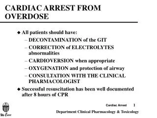
Cardiac Arrest. September 2014 CME. Objectives. Identify the causes of cardiac arrest Identify statistics related to sudden cardiac death Differentiate between sudden cardiac death and cardiac arrest Identify prehospital management for lethal cardiac arrythmias

Cardiac Arrest
E N D
Presentation Transcript
Cardiac Arrest September 2014 CME
Objectives • Identify the causes of cardiac arrest • Identify statistics related to sudden cardiac death • Differentiate between sudden cardiac death and cardiac arrest • Identify prehospital management for lethal cardiac arrythmias • Identify reversible conditions that may contribute to cardiac arrest
Objectives • Identify pathophysiological presentations of cardiac dysrhythmias • Review SMO’s related to cardiac arrest • Review of the components of quality CPR • Discuss “best practices” in cardiac arrest management • Review prehospital use of induced hypothermia for ROSC patients
Cardiac Arrest • 424,000 people experience out-of-hospital cardiac arrest (OHCA) every year • 15-20% annual mortality related to cardiac arrest • 8-10% of all EMS-treated OHCA recover to resume normal lives
Sudden Cardiac Death vs. Cardiac Arrest • Sudden Cardiac Death = “Unexpected death from a cardiovascular cause in a person with or without preexisting heart disease” (Circulation; DOI: 10.1161/CIRCULATIONAHA.111.023838)
Cardiac Arrest = Loss of cardiac function resultant of: • Acute myocardial infarction, OR • Ischemia without infarction, OR • Structural alterations such as scar formation or ventricular dilation secondary to prior infarction or chronic ischemia
What’s the difference…? • It’s a matter of semantics… • The SCD studies focus on mortality rates • Cardiac arrest statistics focus on outcomes; i.e. survival rates • Prehospital providers need to look at the outcomes to determine our level of quality
Risk Factors of Cardiac Arrest • Coronary Heart Disease • Fibrous scar tissue formation on cardiac muscle • Direct effect on pump mechanism and electrical conduction pathways • Ischemia; chronic or acute • Coronary artery blockages • Left ventricular dilation, myocardial stretch • Muscle walls are fatigued • Precursor to congestive heart failure
Risk Factors of Cardiac Arrest • Congestive Heart Failure • Altered calcium regulation • Decreased calcium = less contractile force • Fibrous/Scar formation • Contractile force is inhibited by lack of elasticity of the cardiac muscle • Left ventricular dilation, myocardial stretch • Muscle walls are fatigued • Hypertrophy (left ventricular) • Increase in muscle mass, but the muscle does not increase its pumping ability • In pathological hypertrophy, the heart can increase its mass by up to 150%.
Risk Factors of Cardiac Arrest • Shared Risk Factors • Age • From age 50 to 75 there is an 8x greater risk • Hypertension • Diabetes • Smoking • Obesity • Renal disease • Inflammation • GENETICS!!!!
Genetics and cardiac arrest • Cholesterol • Represents the strongest link genetically • Elevated LDL (the bad one) • Decreased HDL (the good one) • Increases the risk of cardiac arrest at younger ages! • Typically the sub-50 age group; as early as the mid-20’s! • Taken in through food (controllable) • Produced by the body (uncontrollable) • Up to 1000mg/day
Athletes and cardiac arrest • Physical activity can reduce the risk of cardiac arrest…generally • Much media attention given to student athletes with pre-existing cardiac conditions • Helps raise awareness for early cardiac screenings
Pathology of Cardiac Arrest • Heart generally progresses through several cardiac rhythm disburbances…
Cardiac Arrest…Let’s Fix It!! • BLS before ALS! • Must assess before you treat • What’s the most important intervention to be performed in a cardiac arrest??????
CPR “Chain of Survival” • Immediate recognition of cardiac arrest and activation of EMS • Early CPR with emphasis on chest compression • Rapid defibrillation • Effective advanced life support • Integrated post-cardiac arrest care
From the “Cardiac Bible” Circulation: Cardiovascular Quality and Outcomes (AHA Publication; http://circoutcomes.ahajournals.org) • “Summary: Among patients with OHCA, survival from shockable arrhythmias (VT/fibrillation) has improved in recent years after the implementation of guidelines increasing the time devoted to chest compression during resuscitation. These changes include reducing the number of back-to-back rhythm analyses/shocks, eliminating rhythm and pulse checks after each shock, and increasing the ratio of chest compressions to ventilations.”
Breaking it down… “…implementation of guidelines increasing the time devoted to chest compression during resuscitation.” • Minimum 100 compressions per minute • Maximum 120 compressions per minute • Compression IS important, BUT, you must allow FULL RECOIL of the chest to allow the heart to refill with blood to circulate on the next compression
Breaking it down… “…implementation of guidelines increasing the time devoted to chest compression during resuscitation.” • It takes 16 seconds worth of compressions to obtain enough vascular pressure for oxygen exchange to occur within the cells of vital organs • It only takes 3 seconds of no compressions to reduce that pressure back to “0”!!! • Maintain your compressions while you ventilate
Breaking it down… “These changes include reducing the number of back-to-back rhythm analyses/shocks, eliminating rhythm and pulse checks after each shock, and increasing the ratio of chest compressions to ventilations.” • Get on the chest and STAY ON THE CHEST even after defibrillation! • If rhythm converts to a “viable” one after defibrillation STAY ON THE CHEST for one minute before assessing a pulse • When using an AED only stop compressions when the machine tells you to • Ventilate once every 5-6 seconds, or once every 20 compressions…NO MORE
Reality Check… Prehospital personnel are horrible at CPR • Why? • We worry about the “next” intervention • We do too much ALS before BLS • We spend to much time on ET/IV/IO insertions • We think we “already know this”!!!!
Low Frequency…High Intensity • Calendar year 2013 • SCEMSS providers care for more than 30,000 patients • 28 different agencies across almost 300 sq miles • Less than 500 documented, non-traumatic, cardiac arrest patients • That’s 1.6% of total call volume
How many of you could be experts at anything you only practice 1.6% of the time?
Improving CPR outcomes Best Practices • The goal is to save lives • If the rhythm is shockable – Stay and Play • If the rhythm is not-shockable – Load and Go • Only move the patient if… • Provider safety is at risk • The rhythm is not shockable
Improving CPR outcomes Does this mean I may have to stay on scene for 20 minutes doing care? Shouldn’t I be getting this patient to the hospital? • So long as the rhythm is “shockable”, patient survival statistics prove 11% survivability by continuing aggressive CPR and defibrillation. This drops to less than 3% in non-shockable dysrhythmias • What you do in the field is nearly identical to what a physician will do in the hospital. So what’s the hurry when positive patient outcome is the priority?
Improving CPR outcomes “Do unto others…” Knowing what you know now… This V-Fib patient is your spouse, child, parent, sibling, best friend, etc…
Are you still going to halt definitive resuscitation to: • Move them onto a board • Onto the cot • Pile the equipment up • Move through the hallway • Get down/up the stairs • Out to the ambulance • Into the ambulance • Restage your equipment And finally resume quality CPR!!
So we have a survivor! • Induced Therapeutic Hypothermia (ROSC) • Region VII ALS SMO’s, Code 11 • Key points • Cardiac arrest not related to trauma, hemorrhage, or infection • Age >16 • Not currently pregnant • Patient is intubated and unresponsive • Initial temperature > 34 degrees C (93.2 F)
Why ROSC? • American Heart Association • 2005 Updates • Therapeutic hypothermia demonstrates brain cooling in newborn asphyxia improves nuerological outcomes • 2010 Updates • Therapeutic hypothermia in adult cardiac arrest patients shows improved neurological outcome for those that are discharged from the hospital
Why ROSC? • 2010 Updates, cont’d
ROSC • 2010 AHA Outcomes • Cardiac arrest victims that receive therapeutic hypothermia show a 13% survival rate to discharge • ONLY 4% ARE NEUROLOGICALLY INTACT!
ROSC • SMO Algorithm • Return of spontaneous circulation • Initial temp > 34 degrees Celcius • ET in place (NOT A KING AIRWAY DEVICE) • Confirm not responsive to verbal stimuli • Expose patient. Perform 12-lead EKG • Apply ice packs to groin and axilla • Cold saline bolus: 30ml/kg, max 2L • Versed 0.15 mg/kg slow IV push, max 10 mg (for sedation/shivering) with repeat B/P
ROSC • What current science says… • “A new study found that contrary to conventional belief, pre-hospital hypothermia had no effect on the rate of survival to hospital discharge or on neurological outcome among surviving cardiac arrest patients, either among patients with ventricular fibrillation (VF) or non-VF arrest.” • Nuerology Today: Volume 14(2), 16 January 2014, pp 1,9-9
ROSC • From the same study… • “However, the results from Dr. Kim’s trial were not simply that field cooling offered no advantage to patients. Instead, those patients randomized to prehospital cooling experienced re-arrest on the way to the hospital more often – 26 percent versus 21 percent – as well as increased pulmonary edema and use of diuretics.”
ROSC The Decision is Yours… Choose wisely
Why Discuss Cardiac SMO’s? Well, because we are not always following them. • In November and December 2013, only 32% of eligible chest pain patients in SCEMSS received nitroglycerin. • Eligible means appropriate BP, cardiac signs/symptoms, etc…. • In June 2014, only 25% of patients who got Nitro had their BP or pain level properly reassessed after administration. • 2014 to date, SCEMSS medics rarely utilized pacing for symptomatic bradycardia patients. • During most V-Fib calls in the 3rd and 4th Quarter 2013, patients never received lidocaine.
SMO’s: The Template of Care • Template? • “Templates” are used as the basis of creation. We act within our “Medical Orders”; to the degree that it corresponds to how the patient presents • NOT “Guidelines” • Guidelines are “a rule or instruction that shows or tells how something should be done” (http://www.merriam-webster.com/dictionary/guideline)
SMO’s • We already know what to do…we just need to convince ourselves to do it! • Be Confident! Be Proud of Your Knowledge! • SCEMSS encourages providers to advocate for your patients • Use Medical Control to your advantage…NOT as the “Mother May I…” • Keep in mind that an “order” from medical control must be adhered to unless it directly compromises provider or patient safety!
Medical Control Your Friend on the Other End!
Cardiac SMO Review • Let’s review ALS cardiac SMO’s. They are listed on the following slides. • As you go through them, discuss the notes on the slides, as well as any other medications or treatments you have questions about. • ILS/BLS providers – your trimester test will not include ALS SMO’s, but it will include questions on basic cardiac assessment.
Suspected Cardiac SMO • About Aspirin: • We don’t primarily give aspirin to cardiac patients for pain relief. • We give it because aspirin’s blood-thinning properties are linked to better outcomes for cardiac patients. • While aspirin can provide a small amount of pain relief, nitroglycerin and morphine are the true pain-fighters during a heart attack.
Aspirin Cautions • Typical • Known allergy or Hx GI bleeding • Atypical • USE OF BRILINTA • Do not administer ASA to ANY patient on this drug!! • Yes! This is as example of why…“I have a list of medications coming with” is not acceptable! You have to ask for current medications!
Brilinta • BRILINTA is used with aspirin to lower the chance of having a serious problem with heart or blood vessels such as heart attack, stroke, or blood clots • A dose of aspirin higher than 100 mg daily will affect how well BRILINTA works • Doses of ASA higher than 100mg can cause antagonist reactions; including blood clot formation
Suspected Cardiac SMO • Some more points to ponder: • The goal is zero pain. As long as it’s not contraindicated, nitroglycerin is one of the best ways to achieve that goal. • Blood pressure and 1-10 pain levels must be assessed before AND after each administration of nitroglycerin. • IV access is a good idea when giving nitro, in case BP suddenly bottoms out. • And an FYI: • Especially in women and diabetics, weakness, n/v/d, or arm/jaw/back/shoulder pain may be the only symptom of a cardiac event. ALWAYS, do a 12-lead.
Nitro…Or…not to Nitro • For use when… • Suspected cardiac patient • Systolic BP > 110 mmHg • NOT for use when • Patient is taking nitrates for erectile dysfunction • Systolic BP <110 • Patient presents with an Inferior Wall MI on 12-lead EKG
WHOA!!! • Inferior wha’? “Of course I know what the Inferior Wall thing is…It says so on the printout!” Bazinga!
$how Me the $TEMI • Inferior wall STEMI • ST segment elevation is Leads II, III, and aVF • Inferior wall STEMI can effect both sides of the heart – effecting afterload AND preload • Use of NTG may drastically reduce the patients blood pressure to the point of syncope Lead II Lead aVF Lead III