Download
1 / 10
150 likes | 567 Vues
Genetic Collagen Disorders Osteogenesis Imperfecta Ehlers Danlos Syndrome There are others. Types of collagen. Type I (most common): In bone, scar tissue, tendons, ligaments . Type II : Hyaline cartilage .

E N D
Genetic Collagen Disorders Osteogenesis Imperfecta Ehlers Danlos Syndrome There are others.
Types of collagen • Type I (most common): In bone, scar tissue, tendons, ligaments. • Type II: Hyaline cartilage. • Type III: Found in the extracellular matrix of earl granulation (wound healing) tissue; replaced by type I coll. In mature scar tissue. • Type IV: Lens of eye; basal lamina beneath epithelium (esp. in skin, beneath epidermis); capillaries, including glomeruli. • Type V: Interstitial tissue (loose conective tissue); placenta. • List goes up to XXVIII=28…
Collagen Structure Right handed helix of 3 subunits. Each subunit is a LH helix (not an a-helix which is RH). Handednes of a helix: fingers show sense of rotation when travelling in direction that thumb points. Julian Voss-Andreae "Unraveling Collagen“ “a metaphor for aging and growth” http://en.wikipedia.org/wiki/Image:Collagentriplehelix.png
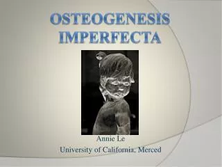
Osteogenesis Imperfecta • General definition: Genetic bone disorder in which bones are weak and fragile. • Various subtypes have been defined more specifically. • Symptoms • Type I (most common, mildest): Bone fragility, blue sclerae. Multiple fractures with minimal cause. • Type II: Severe deformity and bone weakness; death near time of birth due to respiratory insufficiency. • Type III: Progressive skeletal deformity of limbs through childhood, and of spine in late childhood/early adolescence. Sclerae often normal.
Osteogenesis Imperfecta, type III Craig W. Wiesenhutter www.cdaarthritis.com/images_slides
Osteogenesis Imperfecta • Cause • Mutation to collagen gene, usually to alpha subunit of type I collagen gene. Usually autosomal dominant, inherited; occasionally de novo. • Type I: Collagen is relatively normal (by some biochemcial tests) but present in reduced amounts. Due to mutations that affect post-transcriptional- and/or post-translational processing more than the folding of the final product. • Type II: Low amount and low “quality” of collagen. • Type III: Collagen is present in normal amounts, but of insufficient quality. Often due to mutation in gene for alpha subunit of type I collagen which prevents normal triple helix from forming.
Osteogenesis Imperfecta OI type II , type III: mutation in collagen gene → normal collagen triple helix doesn’t form
Ehlers Danlos Syndrome General definition: Joint hypermobility with skin changes Various subtypes have been defined more specifically. Symptoms Unstable, flexible joints (hypermobility) with a tendency to dislocate and subluxate, due to ligaments which are overly stretchable. Elastic, fragile, soft skin that easily forms welts and scars. Intense pain where the joints dislocate is very common. Cause Mutation to collagen gene, usually to type I or type V collagen gene. Autosomal dominant.
Ehlers Danlos Syndrome 20-year-old man presented for consideration of repair of a rectal prolapse that had been present since birth. Physical examination revealed hyperelasticity of the skin (Panel A) with easy bruisability, hypermobility of the joints (Panel B), and a rectal prolapse of about 15 cm in length, which was easily reducible. Results of laboratory tests and chromosome studies were normal. Results of a skin biopsy showed disorganization of collagen bundles in the dermis. Chen and Jao (2007), NEJM 357 (11): e12.
Proteoglycans make extracellular matrix of cartilage a gel Typical proteoglycan in hyaline cartilage is a long hyaluronan (hyaluronic acid, hyaluronate) molecule with attached aggrecan molecules. Aggrecan is itself a proteoglycan, i.e. a protein linked to complex carbohydrates. Chondroitin sulfate, keratan sulfate, heparain, and hyaluronan are glycosaminoglycans. Hyaluronan also found in synovial fluid and vitreous humor. Rooster comb a source for hyaluronan which is key ingredient in “Synvisc”. Aggrecan core protein Hyaluronan backbone with aggrecans attached. Linker protein at base of each aggrecan not shown. (Cell shown here is not to scale: aggrecan molecules are ~50-300 nm long.) Craig W. Wiesenhutter www.cdaarthritis.com/images_slides