Data courtesy: Alex Goddard
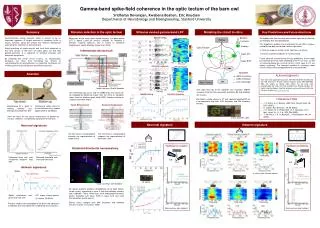
Neuron Chip. Synapses. Neurons. Layer 4/5. E. Retina. AMPA (excitatory). Excitatory. GABA (inhibitory). Layer 8/10. I. Optic Tectum. ACh (cholinergic). Inhibitory. I. I. E. Imc. 40x. 4x. Ipc. Gaze Control. Distractor Suppression. Target Enhancement. ACh. Retina. Retina.

Data courtesy: Alex Goddard
E N D
Presentation Transcript
Neuron Chip Synapses Neurons Layer 4/5 E Retina AMPA (excitatory) Excitatory GABA (inhibitory) Layer 8/10 I Optic Tectum ACh (cholinergic) Inhibitory I I E Imc 40x 4x Ipc Gaze Control Distractor Suppression Target Enhancement ACh Retina Retina I L-4 ACh E L-8 E L-8 ? We hypothesize that neuronal and network signature of attention are linked by ACh neuromodulation This hypothesis predicts that inactivating the Ipc (ACh nucleus) should disrupt both neural and network signatures: • Contrast response function shifts right (less sensitivity) • Gamma-synchrony reduces (if not eliminated) Future work will involve testing the key predictions of the model by inactivating the Ipc, while recording in the OT (in-vivo), as well as microstimulating Ipc (in-vitro) to test if ACh input to OT can induce synchrony. The transient increase in synchrony upon stimulus offset will be incorporated into a revised model. Gamma-band spike-field coherence in the optic tectum of the barn owl Sridharan Devarajan, Kwabena Boahen, Eric Knudsen Departments of Neurobiology and Bioengineering, Stanford University Summary Stimulus selection in the optic tectum Stimulus evoked gamma-band LFP Key Predictions and Future directions Modeling the circuit in-silico Spatial tuning Synchronization among neuronal spikes is known to be an important signature of target selection in primates. Little is known, however, about the cellular and network mechanisms underlying the induction of this synchrony. Using recordings of single neurons and local field potentials in the optic tectum of the barn owl (Tyto alba), we find that gamma-synchrony is a signature of stimulus selection and distractor suppression. By modeling the tectal circuit in-silico, on neuromorphic hardware, we show that mimicking the effects of neuromodulation by acetylcholine is a potential mechanism for evoking synchrony during bottom-up stimulus selection. Being part of the avian gaze control circuitry, the optic tectum (OT) is ideally suited for stimulus selection. Its homolog in primates (superior colliculus, SC) is known to contribute importantly to spatial attention (Muller et al, 2005). LFP spectrogram Isthmotectal microcircuit Contrast response LFP spectrogram Attention Acknowledgments This work was supported by grants NIH1 R01-DC00155-25 (EK) and the NIH Director’s Pioneer Award Program Grant DPI-OD000965 (KB). SD wishes to thank John Arthur for his help with programming the chip, and Alex Goddard and Phyllis Knudsen for kindly sharing images. Spectral analyses were performed with the Chronux toolbox (www.chronux.org) Image courtesy: Phyllis Knudsen ACh input from Ipc to OT facilitates fast excitatory (AMPA) synapses from the retina onto both excitatory (E) and inhibitory (I) neurons. The cholinergic Ipc circuit, and the GABA-ergic Imc circuit can be engaged by bottom-up inputs from the retina or top-down inputs from the forebrain gaze fields (AGF), thereby initiating or suppressing motor output. Spatial tuning Contrast response Literature Cited We model a single column in OT with spatially localized RF on a neuromorphic chip with 1024 excitatory and 256 inhibitory neurons. • J. V. Arthur, K. A. Boahen, IEEE Trans. Neural Netw. 18, 1815 (2007). • H. Luksch, Rev. Neurosci. 14, 85 (2003). • J. R. Muller, M. G. Philiastides, W. T. Newsome, Proc. Natl. Acad. Sci. U. S. A. 102, 524 (2005). • T. Williford, J. H. R. Maunsell, J. Neurophysiol. 96, 40 (2006). Orienting to salient stimuli in the environment (e.g. sudden appearance of a predator) Maintenance of a “goal” in working memory (e.g. distinguishing food from dirt) Here we focus on the neural mechanisms of bottom-up stimulus selection, a fundamental component of attention. Arthur & Boahen, 2007 Neuronal signature Network signature Neuronal signatures The Ipc circuit is well-placed to enhance the representation of target stimuli. The Imc circuit is well-placed to suppress the representation of distractors (red). In-vivo Facilitation of excitation Facilitation of inhibition Detailed isthmotectal neuroanatomy Enhanced firing rate, and sharpened receptive field (RF) Reduced threshold and increased sensitivity Network signatures In collab. with: Shreesh Mysore In-silico Image courtesy: Alex Goddard Ipc (green, biocytin) projects homotopically to the optic tectum (arrow, insert), terminating in layer 5, rich with inhibitory neurons (red, calbindin). These interneurons have widespread horizontal arbors. Excitatory cells (blue, DAPI) in layers 8-10 also send their dendrites up into layer 5. Spikes synchronize and phase lock with LFP LFP shows strong gamma (γ) rhythm (30-90Hz) Retinal axons synapse onto both excitatory and inhibitory neurons in layers 1-5 (Luksch, 2003). Previous models have attempted to link these two signatures of attention, but have ignored the underlying neural circuitry. Data courtesy: Alex Goddard Data courtesy: Alex Goddard