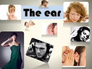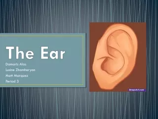
Explore the Intricate Inner Ear System
Discover the detailed structures of the ear, from the external auditory canal to the cochlear duct, explaining mechanisms of hearing and equilibrium. Learn about the dynamic vestibular system and the sensory hair cells in the membranous labyrinth.

Explore the Intricate Inner Ear System
E N D
Presentation Transcript
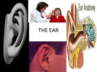
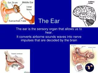
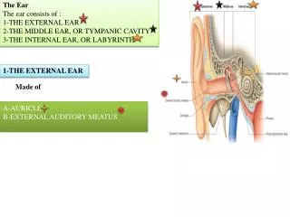
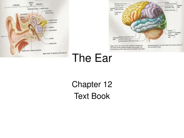
The Ear Otic; Vestibular; Auditory
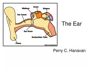
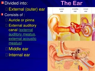
External Ear (contains air); Middle Ear (contains air); Inner Ear (contains fluid) M E I pinna tympanic membrane external auditory canal
Middle Ear: the ossicles Malleus (tympanic membrane) Incus Stapes (oval window) I M S
M I S E. A. Canal TM pharyngotympanic (auditory) tube, connected to nasopharynx region of the throat; functions in equalizing pressure. Normally this tube is flattened and closed, but swallowing or yawning causes it to open temporarily to equalize the pressure of the middle ear cavity with external air pressure. Middle Ear air air AT air
Inner Ear Two layers: Bony labyrinth filled with an aqueous fluid called perilymph. The three subdivisions of the bony labyrinth are the • semicircular canals • vestibule • cochlea. Membranous labyrinth filled with endolymph. This is a second membrane-bound system suspended in the perilymph. The three subdivisions of the membranous labyrinth are: • the semicircular ducts • the Utricle and the Saccule • the cochlear duct.
Inner Ear Semi-circular canals (semi-circular ducts) C U V S Vestibule (utricle & saccule) Cochlea (cochlear duct)
Vestibular System: Dynamic Equilibrium • Semicircular canals: (1) lateral (horizontal), (2) posterior (sagittal), and (3) anterior or superior (frontal) canals. • Contain membranous semicircular ducts, at the base of which is an enlarged region, the ampulla. • Each ampulla contains a crista, consisting of a tuft of hair cells covered with a gelatinous cap, or cupola. • Circular movements of the head move the endolymph in the semicircular ducts, which pushes the cupola in the ampullae, and causes the hair cells to fire impulses, which travel down the vestibulocochlear cranial nerve VIII to the brain.
Inner Ear anterior (frontal) canal Semi-circular canals C U lateral (horizontal) canal V S posterior (sagittal) canal vestibule cochlea
Semi-circular canals contain membranous semi-circular ducts, at the base of which is an enlarged region, the ampulla. Each ampulla contains a crista, consisting of a tuft of hair cells covered with a gelatinous cap, or cupola. ampulla
Hair cells in ampullae Each ampulla contains a crista (hair cell). Circular movements of the head move the endolymph in the semicircular ducts, which pushes the cupola in the ampullae, and causes the hair cells to fire impulses, which travel down the vestibulocochlear cranial nerve VIII to the brain.
Semicircular canals Anterior ‘YES’ Lateral ‘NO’ Posterior TILTINGHEAD
Vestibular System: Static Equilibrium. • The vestibule contains receptors (maculae) that respond to static equilibrium. The maculae respond to gravitational pull, thus providing information on which way is up or down. • The maculae are located in the utricle and saccule. • Maculae consist of hair cells whose cilia project into gelatinous mass containing small grains of calcium carbonate (otoliths). • As the head moves, the otoliths also move, thus triggering nerve impulses in cranial nerve VIII.
Vestibular System: Static Equilibrium A m U m S A maculae
Vestibular System: Static Equilibrium. maculae and otoliths
Vestibular System: Static Equilibrium macular function TILT HEAD BACK
Sense of Hearing • The cochlear duct separates the cochlea into an upper chamber (scala vestibuli) and a lower chamber (scala tympani). The scala vestibuli terminates at the oval window. The scala tympani terminates at the round window. • The cochlear duct supports the organ of corti, which contains the receptors for hearing. These are sensory hair cells resting on the basilar membrane; the cilia of these hair cells project up into the tectorial membrane, and the nerve endings of the cochlear division of the vestibulocochlear cranial nerve VIII, which sends impulses to the auditory association centre in the temporal lobe of the brain.
Cochlear chambers cd td sv cd td
Organ of Corti tm bm tm bm
Summary of the Inner Ear Bony Labryinth (Perilymph) Dynamic Static Hearing Equilibrium Equilibrium Semicircular canals Vestibule Cochlea Oval Window: Scala vestibuli Round Window: Scala tympani Membranous Labryinth (Endolymph) Semicircular ducts Utricle & Saccule Cochlear Duct Ampullae with cristae Maculae Organ of Corti Cupola with hair cells Otoliths Tectorial membrane, hair cells with hair cells Basilar membrane