Download
1 / 41
470 likes | 832 Vues
FUNCTION/DYSFUNCTION OF ENDOCRINE PANCREAS. Diabetes. Anatomy of the pancreas: Both an exocrine and endocrine organ Cells with exocrine function release an alkaline fluid containing sodium bicarbonate and enzymes → pancreatic duct → small intestine

E N D
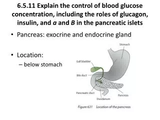
Anatomy of the pancreas: • Both an exocrine and endocrine organ • Cells with exocrine function release an alkaline fluid containing sodium bicarbonate and enzymes → • pancreatic duct → small intestine • Pancreatic “juice” aids in breakdown and digestion of food in the small intestine • Pancreatic exocrine cells = acinar cells
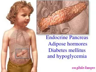
Endocrine Function : • Cells of the Islet of Langerhans synthesize and release hormones into the circulation. • Hormones travel through the bloodstream to target tissues (especially liver and muscle) • At the target cells, hormones bind specific receptors and cause cell changes that control metabolism
Pancreatic endocrine cells regulate carbohydrate, fat, protein metabolism: • Alpha cells – secrete the hormone glucagon (opposite function as insulin) • Beta cells – secrete the hormones insulin and amylin (similar function as insulin) • Delta cells – secrete the hormones gastrin and somatostatin • F cells - secrete hormone pancreatic polypeptide
Beta Cells • Synthesize pre-proinsulin, a protein • This is cleaved by enzymes →proinsulin, then cleaved again → insulin • Insulin is the biologically active hormone that is released into the bloodstream
Insulin secretion is controlled through several mechanisms: • Chemically – high levels of glucose and amino acids in the blood • Hormonally – beta cells are sensitive to several hormones that may inhibit or cause insulin secretion • Neurally – stimulation of the parasympathetic nervous system causes insulin to be secreted.
Insulin secretion is decreased by: • Decreased blood glucose concentration • Increased blood insulin concentration • Sympathetic stimulation
Insulin • Transported through the blood to target tissues where it binds to specific receptors • The binding of insulin to target cells: • Acts as a biochemical signal to the inside of the cell • Overall, cell metabolism is stimulated • There is increased glucose uptake into the cell • Regulation of glucose breakdown within the cell • Regulation of protein and lipid breakdown within the cell
Blood glucose is decreased because insulin causes glucose to leave the bloodstream and enter the metabolizing cells. • With the exception of brain, liver and erythrocytes, tissues require membrane glucose carriers.
Disorder ‑ Diabetes mellitus • The single most common endocrine disorder – group of glucose intolerance disorders • Incidence is estimated at 1-2% of the North American population • Many of these cases are undiagnosed
Diabetes mellitus Historically ‑ distinguished by weight loss, excessive urination, thirst, hunger Excessive urination = polyuria Excessive thirst = polydipsia Excessive hunger = polyphagia Modern characterization is by hyperglycemia and other metabolic disorders
Modern classifications • Type 1 or IDDM ‑ Insulin Dependent Diabetes Mellitus • Type 2 or • NIDDM ‑ Non‑Insulin Dependent Diabetes Mellitus • GDM ‑ Gestational Diabetes Mellitus
Type 1 or IDDM • Accounts for 10% all DM in the Western world ~10-15% have parent or sibling with the disease, Peak age of diagnosis = 12 years • Genetic/environmental/autoimmune factors destroy beta cells • Believed abrupt onset – now immunomarkers and preclinical symptoms have been discovered
Imbalance of hormones produced by islets of Lagerhans : low insulin and high glucagon
Clinical Manifestations: Glucose in urine- Because when insulin is not present, glucose is not taken up out of the blood at the target cells. So blood glucose is very highly increased → increased glucose filtered and excreted in the urine (exceeds transport maximum)
Clinical Manifestations: Weight loss - Patient eats, but nutrients are not taken up by the cells and/or are not metabolized properly Osmotic diuresis results in fluid loss Loss of body tissue by metabolism of fats and proteins
Fats and proteins are metabolized excessively, and byproducts known as ketone bodies are produced. These are released to the bloodstream and cause: Decreased pH (increased acidity), metabolic acidosis Acetone given off in breath
Treatment 1. Administer insulin May be of animal or human origin Cannot be given orally Patient must monitor their blood glucose concentration and administer insulin with the correct timing
2.Control diet • Carbohydrates should make up about 55-60% of patient’s total calories • Fats should make up <30% of patient’s total calories • Proteins should make up about 15-20% of patient’s total calories
3. Monitor exercise • Remember: muscles are a target tissue of insulin, and metabolize much glucose for energy • Sometimes exercise →irregular blood glucose levels So diabetic patients should be monitored when they are exercising
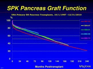
Other: • Pancreatic transplant – so far not successful • Experimental therapies – not as successful as hoped
Type 2 or NIDDM • More common than IDDM, often undiagnosed • It has a slow onset • Most common in those > 40 years, though children are being diagnosed more regularly • May be genetic • Obesity is the greatest risk factor for this disease • And is related to increased incidence in children
NIDDM → insulin resistance in target cells • decreased β cell responsiveness → • Decreased insulin secreted by β cells • Also abnormal amount of glucagon secreted
These effects may be due to: • Abnormally functioning β cells • Decreased β cell mass, or a combination of the two • 3. Target cell resistance to insulin • Due to: • Decreased number of insulin receptors • Postreceptor events may be responsible • Cells “burn out” and become insensitive
Clinical manifestations • Overweight, hyperlipidemia common (but these are precursors, not symptoms) • Recurrent infections • Visual changes, paresthesias, fatigue
Treatment • 1. Weight loss • 2. Appropriate diet (see IDDM above) • 3. Sulfonyl ureas • stimulate βcells to increase insulin secretion • Works only when β cells are still functioning • → An enhancement of insulin’s effect at target cells • 4. Exercise - promotes weight loss
Gestational Diabetes • Due to increased hormone secretion during pregnancy • Seen if patient has predisposition • If previous or potential glucose intolerance has been noted • Important ‑ increased mortality risk for mother, child
Complications of Diabetes Mellitus • Acute: • Hypoglycemia = rapid decrease in plasma glucose = insulin shock • Neurogenic responses – probably due to decreased glucose to hypothalamus. • Symptoms include: • Tachycardia, palpitations, tremor, pallor • Headache, dizziness, confusion • Visual changes
Treatment : provide glucose (I.V. or subcutaneous if unconscious) Observe for relapse
Ketoacidosis – involves a precipitating event: • Increased hormones released w/ trauma increased glucose produced by the body’s cells • This “antagonizes” the effects of any glucose present • Increased ketones in blood • Acid/base imbalance • Polyuria, dehydration Electrolyte disturbances • Hyperventilation (Kussmaul – deep, gasping) • CNS effects • Acetone on breath
Treatment: ‑ low dose insulin Also, administer fluids, electrolytes
Chronic Complications of DM • Neuropathies = nerve dysfunctions → slowing of nerve conduction. • In these patients • Degeneration of neurons →Sensory, motor deficits →Muscle atrophy, paresthesias • G.I. problems, as decreasedmuscle motility • Sexual dysfunction
Microvascular disease – chronic diabetes w/ improper glucose metabolism → thickening of the basement membrane of capillaries, particularly in the eye and the kidney. As the capillary changes in this way, → Decreased tissue perfusion • So ischemia → hypoxia
In the eye – the retina is metabolically quite active, so hypoxia here is a big problem • Retinal ischemia→ • Formation of microaneurisms, hemorrhage, tissue infarct, formation of new vessels, retinal detachment
In the kidney – diabetes is the most common cause of end‑stage renal disease • Injured glomeruli (glomerulosclerosis) • In these patients • Proteinuria (protein is excreted into the urine) → Generalized body edema, hypertension
Macrovascular disease – atherosclerosis Plaque formation increases→ • Increased risk of coronary artery disease, so increased risk of myocardial infarction • Increased risk of congestive heart failure • Stroke • Peripheral vascular disease • diabetic patients face problems with their lower legs and feet • Increased risk of infections