Download
1 / 50
530 likes | 684 Vues
This synopsis explores the historical background, embryology, foetal physiology, histology, and postnatal events of Patent Ductus Arteriosus, emphasizing mechanisms of ductal closure. It covers clinical forms, pathophysiology, and classification of congenital cardiac lesions. The focus is on LR shunts, pulmonary vascular resistance, and the impact on neonatal pulmonary artery pressure. Details on the closure techniques throughout history and the significance of pulmonary blood flow are discussed. This comprehensive overview provides insights into the complexities of PDA.

E N D
Patent Ductus Arteriosus Dr. K. Vanderdonck Cardiothoracic Surgery Charlotte Maxeke Johannesburg Academic Hospital & UNIVERSITY OF THE WITWATERSRAND Hannes Meyer Registrar Symposium 3-5 June 2011
Acyanotic • Cyanotic • LR Shunt • = ↑ PBF • Triad: • FFT • Chest infections • CCF • PDA • ASD • VSD • A-V Canal 2. Obstructive Often asymptomatic Coarctation Aortic stenosis Pulmonary stenosis • 3. Decreased PBF • Cyanosis • Child well, • asymptomatic • Tetralogy • Pulmonary atresia • Tricusp atresia a,b • TGV + PS • 4. Increased PBF • Cyanosis • Triad: • FTT • Chest infections • CCF • Truncus • TAPVC • Tricuspid atresia c • TGV Charateristics Anomalies Classification of Congenital Cardiac Lesions Failure to thrive (FFT); congestive cardiac failure (CCF); pulmonary blood flow (PBF)
Pathophysiology of LR shunts • Clinical importance of pulmonary vascular resistance: • Neonatal pulmonary artery pressure (PAP) greater than that of adults • Reaches adult levels by 2-3 months of age • If PAP remains elevated in presence of a shunt, development of pulmonary vascular obstructive disease (PVOD)
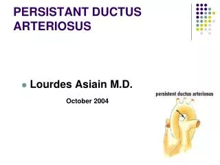
Patent DuctusArteriosusDefinition • Also called ductus of Botalli • Normal vascular structure in foetal life • Extracardiac lesion • Directly connects pulmonary and systemic arterial systems • Persistence of ductus after 3 months in postnatal period abnormal
Patent DuctusArteriosusDefinition • 4 distinct clinical forms: • Isolated PDA in otherwise healthy child • Isolated PDA in premature baby • Associated with more significant cardiac defects • As a life sustaining structure in cyanotic or left-sided obstructive lesions (ductal-dependent)
Patent Ductus Arteriosus Historical Background • Ductus arteriosus & its postnatal closure described by Galen in 131 AD • Physiologic importance of ductus arteriosus elucidated by Harvey in 1628 • 1938 Robert Gross at Boston Children’s Hospital = first successful ligation • 1967 Portsmann used polyvinyl alcohol plug placed with catheter to close PDA • Indomethacin introduced by Heymann in 1976 • 1991 Laborde performed first VATS closure PDA
Patent Ductus Arteriosus Embryology • Derived from distal aspect of the embryological left 6th arch • By 6th week of gestation, ductus arteriosus carries between 55 and 60% of the combined ventricular output
Patent Ductus Arteriosus Embryology • Diverts blood away from high resistance pulmonary circulation to descending aorta and low pressure umbilical placental circulation where gas exchange occurs • Ductal flow directly from PA into descending aorta ductus equal in width to descending aorta and appears as direct extension of PA into descending aorta
Patent Ductus Arteriosus Foetal Physiology • Maintenance of foetalductal patency: • High levels of circulating and locally produced prostaglandins (PGE 2 & PGE 1) • As foetus matures, ductal smooth muscle becomes more sensitive to vasoconstricting effect of pO2, but low pO2 maintains duct patency • pH + other factors play role • RV & LV function in parallel + Share systemic and placental circulations
Patent Ductus Arteriosus Histology • The wall of the ductus differs from the surrounding vascular structures: • Media deficient in elastic fibres • Composed primarily of poorly organized smooth muscle cells in a spiral configuration • Intima thick with increased number of mucoid-filled structures • Smooth muscle sensitive to environmental factors (vasodilating effect of prostaglandins and vasoconstricting effect of pO2)
Patent Ductus Arteriosus Postnatal Events • At birth, rapid circulatory changes • RV & LV function in series • Lung ventilation PVR drops and pulmonary blood flow increases • Due to increased pulmonary venous return, LA pressure rises and PFO closes • PDA closes: • Initially functional and reversible • Later anatomical and irreversible = ligamentum
Patent Ductus Arteriosus Postnatal Ductal Closure • Postnatal closure occurs in 2 stages: • Functional or reversible closure: contraction of medial smooth muscleOccurs within 10-15 hours after birth in full term neonates • Anatomic or irreversible closure: Connective tissue formation with fibrosis produces ligamentum arteriosusCompleted by 2-3 weeks
Patent Ductus Arteriosus Mechanisms of Ductal Closure • Contraction of smooth muscle cells due to: • Increased pO2 following lung ventilation • Decreased PG levels: • Removal placenta = source of circulating PG • Blood flow to lungs removed PG from circulation • Contraction of smooth muscle greatest at pulmonary end, extends to aortic end • Closure may be incomplete at aortic end (ductalampulla or ductal bump)
Patent Ductus Arteriosus Premature Babies • In preterm babies • Overall incidence 30% • Histologically normal ductus but immature • Less sensitive to vasoconstricting effects of pO2, • More sensitive to vasodilating effects of PG • Less likely to respond to postnatal conditions of closure • Trial of Indocid • Early surgery if Indocid fails
Patent Ductus Arteriosus Term Infants • In term infants: • Histology different from normal ductus: • Media contains elastic lamina similar to aortic wall • Smooth muscle organized in fine helocoid spiral fashion • Intima thick with a complete internal elastic lamina • Variable mucoid deposits, lie mostly in media • Is considered a congenital malformation
Patent Ductus Arteriosus Anatomy • PDA = extension of MPA • Curves under the aortic arch • Joins descending aorta at acute angle a few mm beyond origin of LSCA • Recurrent laryngeal nerve curves around PDA • Anatomic variations
Patent Ductus Arteriosus Diagnosis • History • Physical Examination • CXR: Heart Lungs • ECG • Echocardiography (ECHO) + colour Doppler • Often diagnostic of the anatomy • Many operations done on ECHO data only Chest Xray (CXR); Electrocardiogram (ECG); Echocardiography (ECHO)
Patent Ductus Arteriosus Diagnosis • Cardiac catheterization and angiography • To assess PAP + PVR and response to oxygen on pulmonary vasculature • To assess operability • PVR > 8 Wood units in 100% O2 constitutes a contra-indication to surgery ( x 80 to convert to dynes-sec/cm-5 ) • Interventional cardiology • MRI
Patent Ductus Arteriosus Pathophysiology • Dependant on 2 factors: • Size of shunt • Difference between SVR and PVR • At birth, PVR elevated little flow regardless of size • As PVR drops, LtRt shunt increases dependent of size of PDA • Persistent foetal circulation
Patent Ductus Arteriosus Physiological Classification • Physiological Classification: depends • On the size of the PDA • On the degree of pulmonary hypertension and the pulmonary vascular resistance • Important in terms of surgical indication • Classified as small, moderate or large
Patent Ductus Arteriosus Physiological Classification • Small PDA • Qp:Qs < 1.5:1 • Normal PA pressure / normal PVR • Asymptomatic in childhood • Life long risk of infective endocarditis • SBE on PDA − PV − AoV − mycotic aneurysm of descending aorta • Surgery on infected PDA risky • Interventional cardiolgy / transcatheter closure
Patent Ductus Arteriosus Physiological Classification • Moderate size PDA: • Moderate pulmonary hypertension • Do not develop Eisenmenger syndrome • Mild symptoms: some growth retardation, fatigue on effort • May be asymtomatic • Presence of loud murmur with diastolic spillover + thrill
Patent Ductus Arteriosus Physiological Classification • Large PDA • Direct large communication between MPA and Aorta • PA pressure equal to systemic • Qp:Qs increased to a degree dependent on PVR • Can develop Eisenmenger syndrome • CCF – FTT – Chest infections • Systolic murmur
Patent Ductus Arteriosus Physiological Classification • Eisenmenger syndrome: • Severe pulmonary vascular obstructive disease which is irreversible • Presence of suprasystemic PA pressures and PVR with shunt reversal (Rt Lt shunt) • Increasing cyanosis • Death
Patent Ductus Arteriosus Incidence • 5-10% of all congenital cardiac defects • M/F ratio = 1 : 2 • 1 in 1 600 term live births • Incidence higher in preterm babies = 20-30% • Spontaneous closure • Common in premature babies • Rare in term infants
Patent Ductus Arteriosus Incidence • Duct not closing postnatally = pathological • From partial closure to wide open • Factors: • Hypoxia • High altitude • Respiratory distress syndrome • Maternal rubella in 1sttrimestre • Low gestational age • Associated cardiac malformations
Patent Ductus Arteriosus Complications • Death in infancy high due to CCF for large PDA • Death in early, middle adulthood • CCF in moderate size PDA • PVOD + Eisenmenger in large PDA • SBE = complication of small PDA • Respiratory tract infections
Patent Ductus Arteriosus Complications • Ductal aneurysms • Dilatation of the PDA or remaining ductal tissue • Spontaneous or postoperative • Spontaneous = true aneurysms • Postoperative after PDA ligation • Often false aneurysm • Can be true aneurysm
Patent Ductus Arteriosus Complications • Ductal aneurysms • Spontaneous infantile ductal aneurysm • Present at birth or shortly thereafter • Often regress spontaneously • Second type develops in childhood or adulthood • Due to patency at aortic end • Greater tendency for progressive dilatation and rupture
Patent Ductus Arteriosus Treatment • Medical therapy: • Depending on symptoms: • Antifailure treatment • Inotropes • Ventilation • Antibiotics • Pharmacological treatment: Indocid • Surgery or intervention: presence of a duct is an indication for closure, except if pulmonary vascular obstructive disease
Patent Ductus Arteriosus Treatment • Premature babies: • Presence of large PDA associated with organ hypoperfusion + do not tolerate LV overload well • Trial of Indomethacin = inhibitor of prostaglandin synthetase • 0.1 – 0.2 mg / kg 12-24 hourly x 3 doses • Associated with hepatic, renal, platelet dysfunction • Inefficient in term babies
Patent Ductus Arteriosus Surgical Technique • General anesthetic + ventilation • Invasive monitoring • Risk of hypothermia • Patient on right side • Left postero-lateral thoracotomy in 4thintercostal space • Latissimusdorsi incision
Mediastinal pleura opened along descending aorta, to origin of LSCA Superior intercostal vein Care taken to avoid vagus nerve Recurrent laryngeal nerve defines PDA, but don’t go looking for it Patent Ductus Arteriosus Surgical Technique
Patent Ductus Arteriosus Surgical Technique • PDA dissected with blunt angled instrument until completely free
Patent Ductus Arteriosus Surgical Technique Dissect under aorta on both sides PDA
Patent Ductus Arteriosus Surgical Technique Substraction Technique When large PDA + PHT
Patent Ductus Arteriosus Surgical Technique PDA ligation
Patent Ductus Arteriosus Surgical Technique PDA division
Patent Ductus Arteriosus Surgical Technique • Mediastinal pleura is closed: if bleeding, closure will tamponade bleeding and allow exploration • One single pleural drain for 24 hours • In small infants: intercostal muscles approximated with a continuous suture • In older children: 1 or 2 pericostal sutures placed • Patient usually extubatedpostop
Patent Ductus Arteriosus Surgical Technique • In premature baby • Sick: communication with anaesthetist essential • Hand-bagging • Need to release the lung to allow ventilation • Proper dissection essential Clip Single ligation
Patent Ductus Arteriosus Surgical Technique • PDA with severe reversible PHT • PDA with single pulmonary artery • Need cardiac cath and evaluation PVR • Presence PFO • Partial ligation PDA • Restudy later + Interventional closure PDA
Patent Ductus Arteriosus Surgical Technique • Surgery in adult ductus: • More difficult – surgical risk higher than in children • Duct may be calcified • Consider median sternotomy and CPB
Patent Ductus Arteriosus Postoperative Complications • Accidental ligation LPA or aorta in small babies – importance of proper dissection • Recanalisation of ductus: rare even with ligation if properly done • Left vocal cord paralysis – phrenic nerve paralysis uncommon • Chylothorax: rare • Bleeding • Aneurysm of PDA
Patent Ductus Arteriosus Other Therapeutic Modalities • Interventional cardiology • VATS