
Isolation of Phosphorylated Proteins Using Alkaline Phosphatase Mutant
This study focuses on developing a new method for the isolation of phosphorylated proteins using a mutant form of Escherichia coli alkaline phosphatase. The method involves expression, purification, and testing of the mutant enzyme to enhance the efficiency of isolating phosphoproteins in scientific and clinical research.

Isolation of Phosphorylated Proteins Using Alkaline Phosphatase Mutant
E N D
Presentation Transcript
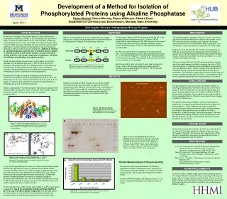
Development of a Method for Isolation of Phosphorylated Proteins using Alkaline Phosphatase Dema Alniemi, Duane Mooney, Royce Wilkinson, Edward Dratz Department of Chemistry and Biochemistry, Montana State University April, 2011 2011 Hughes Scholars Undergraduate Biology Program INTRODUCTION DISCUSSION METHODS Proteomics, the study of the proteins present in cells and their post-translational modifications (PTMs), is an increasingly important field of study. Proteins are responsible for most of the signaling, metabolism, and mechanical action in cells, and PTMs control these activities. More than 400 PTMs have been identified, the most common being covalent attachment of small molecules to intact proteins. One such molecule essential to many protein functions is phosphate. Addition or removal of a phosphate group at specific sites on proteins causes changes in chemical activity or sub-cellular location, thereby controlling cellular activities such as gene expression, metabolism, cell death, and initiation or termination of a protein’s activity. Altered phosphorylation has also been found to play a role in many diseases such as diabetes and cancer. With much of our world’s population being affected with such diseases, work to more efficiently and effectively isolate phosphorylated proteins and to determine changes in phosphorylated proteins in health and disease is becoming increasingly crucial for scientific and clinical applications. My research has been focused on developing a new method for enrichment and isolation of phosphorylated proteins by the use of the Escherichia coli (E. coli ) alkaline phosphatase (ALP) enzyme. ALP is a highly non-specific phosphatase that hydrolyzes the bond between a phosphate group and its attached molecule. Mutant, inactivated ALP has been prepared that retains its ability to bind phosphorylated proteins, but does not take off the phosphate. We plan to use this mutant ALP as an affinity matrix for isolating phosphoproteins. The expression plasmid pEK154 containing either the WT or the mutated gene was transformed into E. coli strain SM547. Transformants were isolated and the DNA sequence of the region of interest was determined by the Nevada Genomics Center, using a sequencing oligo as primer which hybridized starting at nucleotide #95. WT ALP was purified from the periplasm of stationary-phase cells. The crude periplasmic proteins were heat denatured at 80C for 10 min. After centrifugation, the soluble ALP was precipitated in 85% ammonium sulfate. After dialysis, ALP was further purified by ion exchange on Q-sepharose. Enzyme purification was monitored at each step by enzymatic assays, and by SDS polyacrylamide gel analysis. Specific activities were calculated based on moles of PNPP substrate hydrolyzed/min/mg of protein. The desired mutation displayed in Figure 3, S102L, occurred as planned; therefore we were able to work with the mutant form of ALP and perfect the procedures used. However, the mutant enzyme seemed to be lost among the steps of purification, as it was lacking in the proper area as compared to the WT enzyme. Early work led to the idea that the mutation was successful, as DNA was present during the mutagenesis experiment (data not shown), and initial DNA sequencing efforts gave positive mutation results. Activity may have been lost during purification or in a yet undetermined preliminary step. The silver-stained SDS gel (Fig. 5A) demonstrated that osmotic shock and heat treatment markedly increased the purity of ALP. The vast majority of proteins become insoluble when exposed to high temperature, but ALP remains soluble while maintaining its enzymatic activity, making this simple method a major step in purification. Figure 5A also shows that the mutant enzyme was lost or was not well expressed after purification. Further testing needs to be conducted to determine the reasons behind such a result. Mutation of wild type ALP to S102L at active site was created through use of oligonucleotide primers with the desired mutation on the plasmid in a Mutant Strand Synthesis Reaction. 100 101 102 103 104 Wild Type 100 101 102 103 104 Mutant Thr Ser Ala Ala Asp Thr Leu Ala Ala Asp Figure 3. Only a single amino acid change at the active site was made; mutation of nucleotide C371 to T371 leads to the substitution of leucine for serine at amino acid position 102. RESULTS CONCLUSIONS Mutation of the wild type ALP through use of a Mutant Strand Synthesis Reaction was successful, as observed from DNAsequencing results. Methods and protocols used in purification of wild type ALP as well as assays to determine purity and activity were therefore used for both the mutant and wild type ALP simultaneously. Results shown are for mutant and wild type alkaline phosphatase. Transformant # The development of a more powerful and effective method for enriching phosphoproteins promises to be unique in this field of research. Effective methods now work at the peptide level and require protease digestion and “uncoupling” of different PTMs in different parts of proteins. The enriched phosphorylated proteins can be separated intact on 2D gels to more clearly show changes in different PTM isoforms. The ability to better track changes in protein phosphorylation promises to be extremely beneficial for advancing scientific and clinical research. If this method proves to be successful in enriching phosphorylated proteins from their unphosphorylated counterparts, large advances can be made in the field of proteomics, as well as broadly in biomedical research. We have succeeded in developing methods that are foundational for the ultimate goal of this project, and anticipate reaching our target purpose in the near future. 1 2 3 4 5 6 Std Figure 4. Agarose gel showing plasmid DNA with the target gene after mutation. Plasmid supercoils (SC) and nicked circles (NC) are indicated. SC NC A FUTURE WORK Figure 1. Structure of alkaline phosphatase. The wild type E. coli alkaline phosphatase efficiently removes the phosphate from phosphoserine, phosphothreonine and phosphotyrosine sites on proteins. 1 2 3 4 5 6 7 8 9 10 11 12 13 14 15 Future work requires demonstrating a means to covalently bond the mutant ALP to glass beads, providing a simple way to use this reagent. Ultimately, 2D gels and mass spectrometry will be utilized to characterize changes in phosphorylated proteins in health and disease through this method. A B Figure 5-A: Silver stained SDS-PAGE gel of WT and mutant ALP. Lanes from left to right show select steps during increasing salt wash concentration purifications for both WT and mutant forms of the enzyme. Lane #1. WT .05M; #2. WT .075M (1); #3. WT .075M (2); #4 WT .10M (1); #5 WT .10M (2); #6 WT .10M (3); #7 WT .10M (4); #8 MUT .05M; #9 MUT .075M (1); #10 MUT .075M (2); #11 MUT .10M (1); #12 MUT .10M (2); #13.10M (3); #14 MUT .10 (4); #15 Standard REFERENCES Li, W., et al. Development of a universal phosphorylated peptide- binding protein for simultaneous assay of kinases. Biosensors and Bioelectronics, 2009, 24:2871-2877. Tarrant, M., and Cole, P. The Chemical Biology of Protein Phosphorylation. Annual Reviews in Biochemistry, 2009, 78:797-825. Wilkinson, R., and Peters, T. Biochemistry and Molecular Biology Methods, 2010. http://www.chemistry.montana.edu/~martint/ BCHM444/Syllabus_S10.pdf Figure 2. Close-up view of the ALP catalytic site. A: Catalytic center of WT ALP showing interaction between S102 and the phosphate. B: Altered structure of the catalytic center of ALP S102L with a docked phospho-peptide. B Kinetic Measurements of Enzyme Activity Li et al (2009) reported that the S102L ALP mutant binds phosphorylated proteins with high affinity, but fails to remove the phosphate residue. Currently available methods for purifying phosphorylated proteins suffer from various shortcomings. We plan to use ALP(S102L) as a unique means to purify phosphorylated peptides and proteins. Coupled with powerful differential detection methods for revealing changes in phosphorylation developed in our lab, this method is expected to provide insight into the workings of cells as well as provide information as to why certain diseases develop and affect humans. For my research, the mutation S102L was created in ALP by site-directed mutagenesis. Due to the periplasmic location and heat stability of ALP in E. coli, it is easy to purify in high yield. The wild type protein was expressed in E. coli, purified from the periplasmic space, then enzymatic activity and purity were determined as a first major step. ALP enzyme activity was calculated from the rate of appearance at 405nm absorbance due to hydrolysis of p-nitrophenol phosphate (PNPP) to p-nitrophenol (PNP) and phosphate, with an extinction coefficient for PNP of 18,000. The Km of PNPP binding to ALP was found to be 3.1uM which is essentially equivalent to the reported value of 3.6uM. ACKNOWLEDGMENTS I’d like to thank Dr. Edward Dratz and his lab for working with me, with a special thanks to Duane Mooney for guiding me through my project with knowledge, patience, and kindness. Thanks as well to Martha Sellers and the Hughes Undergraduate Biology organization. The Hughes Scholars Program is funded by the Howard Hughes Medical Institute. Figure 5-B. The amount of protein was measured in the indicated salt washes during Q-Sepharose chromatography.


