Protein Structure -Tertiary and Quaternary Structures-
490 likes | 1.1k Vues
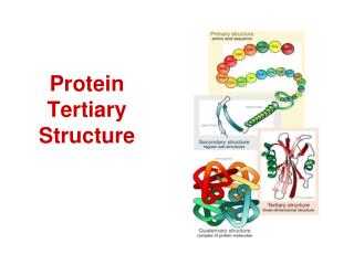
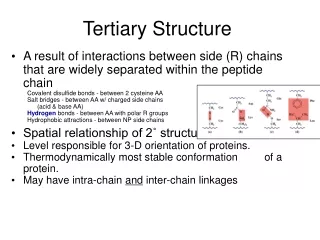
Protein Structure -Tertiary and Quaternary Structures-. Chapter 4 (Page 125-153). 1. Tertiary Structure. 3-D arrangement of all atoms in a protein usually referred to as protein folding. Includes longer-range aspects of AA sequence Interactions of AA between far apart chains

Protein Structure -Tertiary and Quaternary Structures-
E N D
Presentation Transcript
Protein Structure -Tertiary and Quaternary Structures- Chapter 4 (Page 125-153)
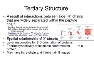
1. Tertiary Structure • 3-D arrangement of all atoms in a protein usually referred to as protein folding. • Includes longer-range aspects of AA sequence • Interactions of AA between far apart chains • Interactions of AA between chains of different secondary structures • For monomeric proteins, tertiary structure is the highest level of structure • Two main types of proteins
1. Tertiary Structure E. Stable, folded conformation dictates the native state of the protein. • Stability defined relative to unfolded state • Native state may NOT be the most stable form of the protein. Often, ligand binding further stabilizes a protein.
1b. Native Proteins are only Marginally Stable • ΔΔG between folded and unfolded = 20 to 65 kJ/mol • Unfolded state is entropically favorable but weak noncovalent interactions within the protein and with water molecules counterbalance this • Water molecules can cause hydrophobic residues to interact in an orderly form • The water molecules adopt a highly structured shell called a solvation layer
2. Fibrous Proteins • Have polypeptide chains arranged in long strands or sheets; not compact • Consist of a single type of secondary structure • Serve the function of providing support, shape, and external protection • The strength of fibrous proteins is enhanced by covalent cross-links between polypeptide chains, such as disulfide bonds
2I. α-Keratin • Displays strength and structural function • Found in hair, nails, skin • Part of a broader family of proteins called intermediate filament (IF) proteins.
2I. α-Keratin D. All IF proteins share structural features exhibited by α-Keratin • Consist of right-handed α-helices in a left-handed coiled coil (see structure in hair) • Parallel orientation of the strands • ~5.2 Å per turn
2I. α-Keratin • The surfaces where the two α-helices touch are made up of hydrophobic AA residues (Namely Ala, Val, Leu, Ile, Met, and Phe) • The two-chain coiled coils combine in higher-order structures called protofilaments and protofibrils stabilized by disulfide bonds
2II. Collagen • Provides strength • Found in connective tissue such as tendons, cartilage, bones • Helical (α-chain) but unlike α-helix • Left-handed • 3 AA per turn • High Gly content D. Three α-chains are supertwisted in a right-handed manner E. The triple helix has higher tensile strength than a steel wire of equal cross section
2II. Collagen F. Many triple-helices assemble into a collagen fibril • Collagen molecules alligned in a staggered fashion • Cross-linked by unusual type of covalent bonds involving Lys, His, or the uncommon AA 5-hydroxylysine
3. Globular Proteins • Consist of different segments of a polypeptide chain that fold into a compact structure i.e. Human Serum Albumin (64.5 kDa; 585 AA) Native Globular form: 100 x 60 Å βconformation: 2000 x 5 Å αHelix: 900 x 11 Å B. Include enzymes, transport proteins, motor proteins, regulatory proteins, immunoglobulins
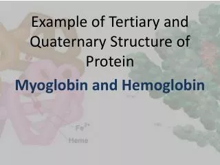
3I. Myoglobin • 3D structure determined from the x-ray diffraction studies of John Kendrew et al (1950s) • Relatively small; 16.7 kDa; 153 AA • Oxygen-binding protein of muscle cells • Distributes and stores O2 in muscle D. Consist of 8 αhelices and 8βturns E. Fe(II) bound heme prosthetic group, responsible for binding O2
3I. Myoglobin Heme: Porphyrin Ligand Covalent bond with His residue • Reversible binding of O2 to Fe(II) is important for breathing. • Fe(II) oxidation to Fe(III) results in dissociation of O2because Fe(III) has a poor affinity for O2.
3I. Myoglobin F. Much of the protein’s stability is derived from hydrophobic interactions. • The hydrophobic R groups are in the interior • Only 4 H2O molecules in the interior • The dense hydrophobic core is typical of globular proteins G. Polar R groups are mostly on the surface surrounded by water Red: Hydrophilic AA Blue: Hydrophobic AA Green: Heme
3II. Protein Folding Protein folding can be quite complex and therefore it is easiest to discuss it in terms of common structural patterns. Supersecondary structures (i.e. motifs or folds) are particularly stable arrangements of several elements of secondary structure and the connections between them. Will discuss them in the context of protein folding rules.
3II. Protein Folding • Hydrophobic interactions are extremely important to protein structure stability. The burial of hydrophobic AA (to exclude water) requires at least two layers of secondary structure. B. αhelices and βsheets are generally in different structural layers due to inefficient hydrogen bonding
3II. Protein Folding C. Polypeptide segments adjacent to each other in the primary sequence are usually stacked adjacent to each other in the folded structure D. Connections between elements of secondary structure can not cross or form knots
3II. Protein Folding E. The βconformation is most stable when the individual segments are twisted slightly in a right-handed sense. • Twisting leads to twisted segments TwistedβSheet βBarrel
3IIc. Organization of Proteins based on Motifs • All α • All β Serum Albumin Bacterioferritin α-Amylase inhibitor tendamist UDP N-acetylglucosamine acyltransferase
3IIc. Organization of Proteins based on Motifs C. α/β(The α and βsegments are interspersed) D. α + β(The α and βsegments are segregated) Alcohol Dehydrogenase Pilin
3III. Tertiary Structure and Evolutionary Ties • Protein tertiary structure is often more reliably conserved than primary sequence. • Protein structure can give great insight into evolution • Proteins with significant primary sequence similarity and/or significantly similar function are grouped into the same Protein Family • Two or more families with little primary sequence similarity but same major structural motifs and similar functions are grouped as Super Families
4. Quaternary Structure • When a protein has two or more polypeptide subunits held together by hydrophobic interactions, hydrogen bonds, disulfide bonds • Is the highest level of structure for this type of protein • Multisubunit proteins tend to play regulatory roles • Binding of small molecules may affect the interaction between subunits • Separate subunits may play different but related functions
4I. Nomenclature • A multisubunit protein is called a multimer and can be named by the number of subunits • If 4 subunits, tetramer • Multimer with just a few subunits (≤ 4) is called an oligomer • A repeating structural unit, whether a single subunit or a group of subunits, is called a protomer
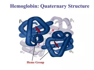
Hemoglobin is a Tetramer • 4 polypeptide chains • 4 Fe(II)-heme groups for O2 binding • 2 α, 2βsegments Also known as (AKA) Dimer of αβprotomers α Subunits β Subunits
4Ib. Geometry • A multimer may be composed of several nonidentical subunits resulting in complex, asymmetric structure • Typically though, multimers are arranged in one or a limited set of symmetric patterns • Rotational symmetry- Individual subunits can be superimposed on others by rotation about one or more rotational axes • Helical symmetry- The units are related by a screw axis (a translation and rotation operation)
4Ic. Rotational Symmetry • Cyclic Symmetry- Rotation about a single axis. • The protein has a symmetry defined by convention as Cn C: Cyclic n: # of subunits related by the axis
4Ic. Rotational Symmetry B. Dihedral Symmetry- A twofold rotational axis intersects a n-fold axis at right angles • Dn: Protein has 2n protomers i.e. D2 protein has 4 protomers
4Ic. Helical Symmetry • Proteins with helical symmetry form structures that are open-ended and spiral i.e. Capsid: Protein shell of a virus Capsid Helical Symmetry
5. Protein Stability • Protein Size • Proteins subunits are typically < 100 kDa • Size of proteins is limited by the error frequency during protein biosynthesis • Error frequency: 1 mistake in 10,000 AA • Though low, the mistake has high probability for production of damaged, unstable protein • Protein can’t fold • Important residue for binding is missing or a conservative substitution did not occur
5. Protein Stability B. Protein Cleavage and Folding • Protein subunits (>100 AA) often fold into two or more stable, globular units called domains. • A domain will often retain 3D structure even when separated from large protein via cleavage C. Protein Denaturation and Folding • Denaturation is a loss of 3D structure, which results in the loss of function • Complete unfolding does not occur nor complete randomization of conformation
5I. Sources of Denaturation • Heat: Thermal denaturation can be gauged by a number of techniques, such as fluorescence. Changes in environment of aromatic AA, changes protein fluorescence. Unfolding exposes Trp residues and increases fluorescence. Unfolding, Fluorescence
5I. Sources of Denaturation • Heat affects the weak interactions in a protein in a complex manner • If increase Temp. slowly, a protein’s conformation remains intact until an abrupt loss of structure occurs over a narrow temp. range. • The midpoint of this range is Tm (Melting temp.) • Abruptness indicates there is cooperativity in the unfolding process. Loss of structure in one part leads to instability in another. • If not cooperative, protein unfolding would occur just by raising the Temp. Would be a linear dependence.
5I. Sources of Denaturation • Heat denaturation can be reversible, semi reversible, or irreversible • Classic irreversible example Albumin
5I. Sources of Denaturation B. Extreme pH alters net protein charge • Changing protonation states can disrupt hydrogen bonds C. Organic solvents D. Solutes such as urea and guanidine hydrochloride (Gu·HCl) E. Detergents Note: C, D, E disturb hydrophobic interactions F. Reducing agents • Break up disulfide bonds
5II. Protein Renaturation Certain proteins will regain their native structure and their biological activity if returned to conditions in which the native conformation is stable
5III. Models for Protein Folding The “correct” AA primary sequence is necessary for proper tertiary structure folding. (Christian Anfinsen, 1950s). Protein folding is NOT RANDOM. • Hierarchical Fold • Local secondary structure first • Longer range supersecondary structures next • Tertiary then quaternary structure
5III. Models for Protein Folding B. Collapsed State Model (Molten globule) • Folding is initiated by a spontaneous collapse of the polypeptide into a compact state mediated by hydrophobic interactions
5IV. Folding facilitated by specialized proteins Folding for many proteins is facilitated by the action of specialized proteins. • Molecular chaperones are proteins that interact with partially folded or improperly folded polypeptides • Hsp70 (MW 70 kDa) family of proteins • Chaperonins • Two enzymes catalyze isomerization reactions • Protein disulfide isomerization (PDI) • Peptide prolylcis-trans isomerase (PPI)
6. Techniques to Study Protein Fold • X-ray Crystallography Steps needed • Purify the protein • Crystallize the protein • Collect diffraction data • Calculate electron density • Fit residues into density Pros • No size limits • Well-established Cons • Difficult for membrane proteins • Cannot see hydrogens
6. Techniques to Study Protein Fold B. Nuclear Magnetic Resonance (NMR) Steps needed • Purify the protein • Dissolve the protein • Collect NMR data (Typically 2D 1H NMR) • Assign NMR signals • Calculate the structure Pros • No need to crystallize the protein • Can see many hydrogens • Can study proteins in motion Cons • Difficult for insoluble proteins • Works best with small proteins