PARTS
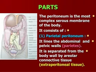
PARTS. The peritoneum is the most complex serous membrane of the body. It consists of : (1) Parietal peritoneum : It lines the abdominal and pelvic walls (parietes). It is separated from the body wall by areolar connective tissue (extraperitoneal tissue). PARIETAL PERITONEUM.


PARTS
E N D
Presentation Transcript
PARTS • The peritoneum is the most complex serous membrane of the body. • It consists of : • (1) Parietal peritoneum : • It lines the abdominal and pelvic walls (parietes). • It is separated from the body wall by areolar connectivetissue(extraperitoneal tissue).
PARIETAL PERITONEUM • It varies in different regions. • It contains a large amount of fat in the area of the kidney.
(2) VISCERAL PERITONEUM • It is the reflection of the parietal peritoneum on the organs. • It is firmly united to the viscera which it covers.
PERITONEAL CAVITY • It is the potential space between the parietal and visceral layers. • It consists of : • A. Greater sac : • It is the main region. • B. Lesser sac (omental bursa) : • It is a diverticulum behind the stomach.
PERITONEAL CAVITY • The lesser omentum separates the greater and lesser sacs. • The two sacs communicate through the opening of the lesser sac (Epiploic foramen).
PERITONEAL CAVITY • In male, the cavity is a closed sac.
PERITONEAL CAVITY • In female, it communicates with the exterior through the uterine tubes, uterus and vagina.
PERITONEAL FLUID • It is secreted by the peritoneum. • It contains leukocytes. • It ensures that the mobile viscera glide easily on each other. • It is not static. • Its accumulation causes ascites.
INTRA PERITONEAL ORGANS • They are organs which are totally covered with visceral peritoneum. • They are not actually within the peritoneal cavity. • Stomach. • Spleen. • Jujenum and Ileum.
RETROPERITONEAL ORGANS • They are organs which are partially covered with visceral peritoneum. • Ascending and Descending colon. • Kidneys. • Pancreas.
PERITONEAL LIGAMENTS • They are two- layered folds of peritoneum. • They connect the solid viscera to the abdominal wall. • In the LIVER, it is connected to the diaphragm by: • Falciform ligament. • Coronary ligament. • Right and Left triangular ligaments.
OMENTA • They are two layered folds of peritoneum. • They connect the stomach to another viscus. • 1. Greater omentum. • (2) Lesser omentum.
GREATER OMENTUM • It connects the greater curvature of the stomach to the transverse colon. • It hangs down like an apron to cover the coils of small intestine. • It folded back on itself to be attached to the transverse colon.
LESSER OMENTUM • It extends between the lesser curvature of the stomach and the under surface of the liver (porta hepatis and fissure for ligamentum venosum) .
GASTROSPLENIC LIGAMENT (OMENTUM) • It connects the stomach to the hilum of the spleen.
SPLENICORENAL LIGAMENT • It connects the hilum of the spleen to the left kidney.
MESENTERIES • They are two layered folds of peritoneum. • They connect parts of the intestine to the posterior abdominal wall. • Mesentery of small intestine. • Transverse mesocolon.
CONTENTS • Blood vessels. • Lymph vessels. • Nerves. • Fat.
SAGITTAL SECTION OF PERITONEUM • 0. Parietal peritoneum lining the anterior abdominal wall ascends upwards to the undersurface of the diaphragm.
SAGITTAL SECTION OF PERITONEUM • 0. It is reflected onto the upper surface of the liver to the left of the falciform ligament. • It forms the anterior layer of the left triangular ligament.
PERITONEUM (SAGITTAL SECTION) • 0. It covers the anterior and inferior surfaces of the liver until it reaches the porta hepatis. • 0. It passes to the lesser curvature of the stomach as the anterior layer of the lesser omentum.
PERITONEUM (SAGITTAL SECTION) • 0. It covers the anterior surface of the stomach. • 0. It leaves the greater curvature to form the anterior layer of the greater omentum.
PERITONEUM (SAGITTAL SECTION) • 0. The greater omentum forms a fold in front of the coils of intestine. • 0. On reaching its lowest limit, the peritoneum folds on itself to form the posterior layer of the greater omentum.
PERITONEUM (SAGITTAL SECTION) • 0. The peritoneum forms the posterior layer of the transverse mesocolon. • 0. It passes to the anterior border of the pancreas and the 3rd part of the duodenum.
PERITONEUM (SAGITTAL SECTION) • 0. The peritoneum leaves the posterior abdominal wall as the anterior layer of the mesentery of the small intestine.
PERITONEUM (S.S) IN FEMALE • It descends down to cover the anterior surface of the rectum. • It is reflected onto the posterior surface of the upper part of the vagina and forms the rectouterine pouch (pouch of Douglas).
PERITONEUM (S.S) IN FEMALE • It passes over the upper surface of the uterus and reflected from its anterior surface onto the upper surface of the bladder. • It forms the shallow uterovesical pouch. • It passes from the bladder onto the anterior abdominal wall.
PERITONEUM (S.S) IN MALE • From the anterior surface of the rectum, it reflected onto the upper part of the posterior surface of the bladder and the seminalvesicles. • It forms the rectovesicalpouch. • It passes from the bladder onto the anterior abdominal wall.
TRANSVERSE SECTION OFPERITONEUM AT (L4) • The parietal peritoneum lining the anterior abdominal wall is mostly smooth. • It is raised by two ridges : • Median umbilical ligament (urachus) : remains of fetal allantois. • Lateral umbilical ligaments : obliterated umbilical arteries.
T. S. OFPERITONEUM AT (L4) • It ascends onto the posterior abdominal wall and becomes continuous with the visceral peritoneum which covers the front and sides of the ascending and descending colon.
T. S. OF PERITONEUM AT (L4) • Para colic gutters: (grooves) lie medial and lateral to the ascending and descending colons.
T. S. E OFPERITONEUMAT (L4) • At the level of the aorta and inferior vena cava, it becomes continuous with mesentery of the small intestine.
T. S. OF PERITONEUM AT (T12) • 1. The parietal peritoneum above the umbilicus forms • The Falciformligament.
FALCIFORM LIGAMENT • It is a sickle – shaped fold. • It connects the anterior surface of the liver to the diaphragm and anterior abdominal wall. • Its free border contains ligamentum teres (obliterated umbilical vein).
T. S. OF PERITONEUMAT (T12) • 2. At the left side of the abdomen : • 0. The parietal peritoneum becomes continuous with the visceral peritoneum covering the lateral margin and anterior surface of left kidney.
T.S. OFPERITONEUMAT(T12) • 0. It passes to the hilum of the spleen as the posterior layer of the splenicorenal ligament. • 0. It covers the spleen. • 0. It is reflected at the hilum as the anterior layer of the gastrosplenicligament.
T. S. OF PERITONEUM AT (T12) • 0. It covers the anterior surface of the stomach . • 0. It leaves the lesser curvature as the anterior layer of the lesser omentum. • 0. On the right, the lesser omentum has a free border.
T. S. OF PERITONEUM AT(T12) • 0. The peritoneum is folded around the bile duct, hepatic artery and portal vein. • 0. It forms the posterior wall of the lesser omentum (anterior wall of the lesser sac).
T. S. OF PERITONEUM AT(T12) • 0. It leaves the greater curvature of the stomach. • 0. It forms the posterior layer of the gastrosplenic ligament. • 0. At the hilum of the spleen, it is reflected as the anterior layer of the splenicorenal ligament.
T. S. OF PERITONEUM AT (T12) • 0. It covers the anterior surface of Aorta, Inferior venacava and Pancreas. • 0. It forms the posterior wall of the lesser sac. • 0. It passes onto the anterior surface of the right kidney.
T. S. OF PERITONEUM AT (T12) • 0. It passes onto the anterior surface of the right kidney. • 0. It sweeps around the lateral abdominal wall to reach the anterior abdominal wall. • 0. It forms a continuous layer around the abdomen.
LESSER SAC • It is a peritoneal pouch. • Position : • Behind the lesser omentum and stomach. • In front of the structures of the posterior abdominal wall (pancreas & duodenum).
LESSER SAC (EXTENSIONS) • Superior recess : • As far as the diaphragm & caudate lobe of the liver. • Inferior recess : • Between the layers of the greater omentum. • This is often obliterated.
LESSER SAC (BOUNDARIES) • 1. Left : • Spleen. • Gastrosplenic & Splenicorenal ligaments. • 2. Right : • Opening of the lesser sac (epiploic foramen). • 3. Below: • Left free border of the greater omentum.
EPIPLOIC FORAMEN Anterior : • Free border of the lesser omentum with its contents (bileduct, hepatic artery and portal vein).
EPIPLOIC FORAMEN • Posterior : • Inferior vena cava.
EPIPLOIC FORAMEN • Superior : • Caudate lobe (caudate process) of the liver. • Inferior : • First part of the duodenum.
GREATER SAC • It is divided by the transverse mesocolon into : • (1) Supra colic part. • (2) Infracolic part.
SUPRA COLIC PART • It contains : • 1. Anterior subphrenic between the liver and diaphragm. • This is divided by the falciform ligament into: • A. Right anterior subphrenic. • B. Left anterior subphrenic.
SUPRA COLIC PART • 2. Right posterior subphrenic(hepatorenal) pouch. • It is between the right lobe of the liver, right kidney and right colic flexure. • 3. Right extraperitoneal space : • Between the layers of the coronary ligament (barearea) of liver.