Download
1 / 65
670 likes | 953 Vues
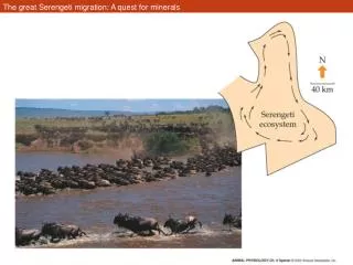
The great Serengeti migration: A quest for minerals. Digestive system. Functions Organs. Organs of alimentary canal. Figure 23.2. Month Esophagus Stomach Small intestine Large intestine Accessory organs Salivary glands, liver, pancreas, gall bladder. Figure 23.1.

E N D
Digestive system • Functions • Organs
Organs of alimentary canal Figure 23.2
MonthEsophagusStomachSmall intestineLarge intestineAccessory organsSalivary glands, liver, pancreas, gall bladder Figure 23.1
Nutrition • Proteins • Lipids • Carbohydrates • Vitamins and minerals
Feeding Examples of feeding adaptations Food chains
Figure 4.6 Some species feed by targeting and subduing individual food items (Part 1)
Figure 4.8 Specialization of an invertebrate feeding apparatus (Part 1)
Figure 4.8 Specialization of an invertebrate feeding apparatus (Part 2)
Figure 4.12 Reef-building corals of warm waters need light because they are symbiotic with algae (2)
Figure 4.9 Short food chains deplete energy less than long food chains do
Digestive systems of insects and crustaceans • Crustaceans’ digestive system is separate from the excretory system • Insects– the Malpighian tubules – excretory system is connected at the junction of the midgut and hindgut
Figure 4.16 The digestive systems of two types of arthropods: insects and crustaceans
Stomach (continued) • Contractions of the stomach churn chyme. • Mix chyme with gastric secretions. • Push food into intestine. Insert fig. 18.5
Small Intestine • Each villus is a fold in the mucosa. • Covered with columnar epithelial cells interspersed with goblet cells. • Epithelial cells at the tips of villi are exfoliated and replaced by mitosis in crypt of Lieberkuhn. • Lamina propria contain lymphocytes, capillaries, and central lacteal. Insert fig. 18.12
Histology of the Alimentary Canal Figure 23.6
Sensors of the GI tract– regulatory mechanisms • Mechanoreceptors and chemoreceptors involved • Located in the walls of the tract organs • Sensors respond to • Stretching • Osmolarity • pH • Presence of substrates and end-products
Regulatory mechanisms (2) • Receptors initiate reflexes • Activate of inhibit glands that secrete digestive juices • Stimulate smooth muscle of GI tract • Move food along the tract • Mix lumen content
Peristalsis and Segmentation Figure 23.3
Adaptation associated with animal’s diet • Microbe-assisted digestion –animals in hydrothermal vents-trophosomes • Dentition/mouth parts • Length of digestive tract • Herbivores • Carnivores • Omnivores • Sharks • Birds
Microbe-dependent digestion • Digestion assisted by microbes
Animals maintain symbiosis with three categories of microbes • Heterotrophic microbes • Organic compounds of external origin • Autotrophic microbes • Synthesize organic molecules from inorganic precursors • Chemosynthetic • Photosynthetic
Figure 4.13 Hydrothermal-vent worms are symbiotic with chemoautotrophic bacteria (Part 1)
Hydrothermal-vent worms • Symbiotic with chemoautotrophic bacteria- trophosomes • Worms have not mouth, gut, or anus • Food comes from sulfur-oxidizing chemoautotrophic bacteria • Organic molecules from bacteria meets nutritional needs • Vents- source of H2S
Hydrothermal-vent worms • Symbiotic with chemoautotrophic bacteria- trophosomes • Worms have not mouth, gut, or anus • Food comes from sulfur-oxidizing chemoautotrophic bacteria • Organic molecules from bacteria meets nutritional needs • Vents- source of H2S
Figure 4.13 Hydrothermal-vent worms are symbiotic with chemoautotrophic bacteria (Part 2)
Comparison of the digestive tracts of carnivores and herbivores • Carnivores- foregut digestion • Herbivores • Hindgut • Foregut
Stomach of ruminants • Several chambers • Rumen – first chamber/fermentation occurs • Regurgitate fermenting materials from the rumen into mouth • Further grinding and reswallow • From rumen reticulum omasum abomasum (true stomach)
Functions of microbes in ruminants • Synthesize B vitamins, essential amino acids • Fermentative breakdown of compounds that animals cannot digest– cellulose • Recycle waste nitrogen from animal metabolism • Make ammonia so other microbes can use it as nitrogen source
Hind and midgut fermenters • Enlarged cecum/colon • Rabbits, horses, zebras, rhinos, apes, elephants • Break down of cellulose and carbohydrates • Forms short-chain fatty acid • B vitamins- not utilized, lost in feces • Coprophagy– rabbits eat special soft feces
A comparison of the digestive tracts of a carnivore (coyote) and a herbivore (koala)
Digestion and absorption • Digestive enzymes in 3 spatial contexts • Intraluminal enzymes • Membrane-associated enzymes • Intracellular enzymes
Intracellular and extracellular digestion • Intraluminal and membrane-associated enzymes are responsible for extracellular digestion • Intracellular enzymes are responsible for intracellular digestion • Advantages and disadvantages of intra- and extracellular digestions?