Download
1 / 47
490 likes | 992 Vues
The Anti-carcinogenic Effect of Echinacea purpurea and Echinacea pallida on BT-549. Nicole Driggins, Dr. E. Lewis Myles, & Dr. Todd Gary Department of Biological Sciences & Center of Excellence Tennessee State University, Nashville TN.

E N D
The Anti-carcinogenic Effectof Echinaceapurpurea and Echinaceapallidaon BT-549 Nicole Driggins, Dr. E. Lewis Myles, & Dr. Todd Gary Department of Biological Sciences & Center of Excellence Tennessee State University, Nashville TN
Medicinal plants have been used to treat different ailments for centuries. Salicin – willow tree Medicinal Plants
Taxol® (Paclitaxel) • In 1975 researchers discovered that taxol, a substance found in the bark of a yew tree, decreases the production of cancerous tumors. • In 1994, FDA approved the use of taxol to treat breast cancer.
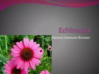
Echinacea • Genus: Echinacea • Family: Asteraceae • Number of species: Nine • North American Plant • Medicinal plant noted for the use of stimulating the immune system.
E. angustifolia* E. atrorubens E. laevigata** E. pallida* E. paradoxa E. sanguinea E. simulata E. tennesseensis** E. purpurea* Species of Echinacea
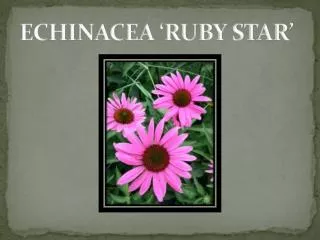
Figure 3. Echinacea purpurea Characterized by the white flowering rays that reflex backwards.
Figure 4. Echinacea pallida Characterized by long, branching, flowering stems and slender purple ray flowers.
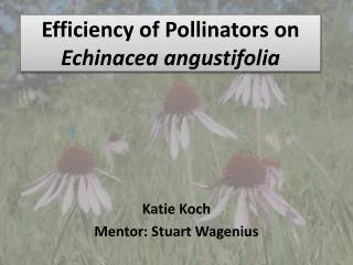
Figure 6. Echinacea augustifolia Characterized by the purpleflowering rays that reflex backwards.
Figure 7. Echinacea tennesseensis Its rays spread up and outward to form an inverted cup-shaped corolla.
Materials: Part One • Plantextract • Breast Cancer Cell Line • 50 ml Tissue Culture Flasks • Media • PBS • Control: DMSO • Six Well Culture Plate • Hemocytometer • Trypan Blue
Breast Cancer Cells • BT-549 cell line (isolated in 1978) • The cells are from a 72 year old Caucasian female. • Grown & maintained in the lab.
Growth Media • RPMI – 1640 • Developed at Roswell Park Memorial Institute in 1966 for the growth of human leukemia cells. • Demonstrated a universal use in the growth and support of several mammalian cells that are anchorage dependent.
PBS • Phosphate Buffered Saline • (Na2HPO4·7H2O, NaH2PO4·1H2O • NaCl) • Isotonic solution used to wash • the cancer cells. • Non-toxic to the cancer cells
DMSO • Dimethyl Sulfoxide • Used to dissolve components of the plant • Non-toxic to the cancer cells • Obtained from Sigma-Aldrich
Method • The cells are removed from the tissue flasks. • The cells are counted by using a hemocytometer. • The same number of cells are placed in each well of the culture flask, which contains media.
Figure 8.Six Well Cell Culture Plate Ech. 1 DMSO 1 Ech. 2 DMSO 2 Ech. 3 DMSO 3
Anti-Carcinogen Procedure Media + Cancer Cells + Plant Extract or Control + 4 Day Incubation + Remove and Count Viable Cells
Plantextract Breast Cancer Cell Line 50 ml Tissue Culture Flasks Media Hemocytometer 20 ml Tissue Culture Flasks PBS Control: DMSO Cell Culture Slides Apoptosis Kit Confocal Microscope Materials: Part Two
Apoptosis Assay • Expose cells to E. purpurea or E. pallida for two days • Expose cells to DMSO for two days • Expose cells to labeling dye • Observe under confocal microscope • Viable Cells = green • Necrotic Cells = red & green • Apoptotic Cells = red • Sigma-Aldrich
Figure 15.The Mechanism of a Confocal Microscopehttp://www.physics.emory.edu/~weeks/confocal/
Fig. 18 Apoptosis FADD
Fig. 19 Apoptosis Cont.’d FADD Pro-caspase 8
Fig. 20 Apoptosis Cont.’d FADD Bid Caspase 8
Fig. 21 Apoptosis Cont.’d Bid Bax Mitochondrion Cytochrome C
Fig. 22 Apoptosis Cont.’d Apaf -1 Pro-caspase 9 Cytochrome C
Fig. 23 Apoptosis Cont.’d Apaf-1 Caspase Cascade Caspase 9 Cytochrome C Apoptosis
Figure 24.Apoptosis Cont.’d Bax Mitochondrion FADD Caspase 8 Bid Apaf-1 Caspase Cascade Caspase 9 Cytochrome C Apoptosis
Conclusion • Further Apoptosis Analysis with E. purpurea • Further Apoptosis Analysis with DMSO • Further Apoptosis Analysis with E. pallida • Determination of E. purpurea and E. pallida component(s) that causes the effect on the breast cancer cells • Addition of a normal breast cell line and an African-American breast cancer cell line • Western Blot Analysis
Acknowledgements • NASA, NIH • Dr. E. Lewis Myles • Dr. Todd Gary • Dr. Johnson, Dr. Blackshear, Dr. Kahlon and Dr. Whalen • Mrs. Yvonne Myles • Dr. John Robinson, Ryan Johnson, Brad Smith • Wendel Forston, Clifton Randell, Tanisha Taylor, Alicia Cleveland, Tim Udoji, Chasity Bradley and Tamela Hunt
References • 1. Goldhaber-Fiebert, S., Kemper, K.J. [1999]. Echinacea: E. angustifolia, E. pallida, and E. purpurea. The Center for Holistic Pediatric Education and Research. http://www.mcp.edu/herbal/default.htm • 2. Letchamo, W., Livesey, J., Arnason, T.J., Bergeron, C.,and Krutilina, V.S. [1999]. Cichoric Acid and Isobutylamide Content in Echinacea purpurea as Influenced by Flower Developmental Stages. p. 484-498. In: J. Janick [ed.], Perspectives on New Crops and New Uses. ASHS Press, Alexandria, V.A. • 3. McKeown, K.A. [1999]. A Review of the Taxonomy of the Genus Echinacea. p. 482-489. In: J. Janick [ed.], Perspectives on New Crops and New Uses. ASHS Press, Alexandria, V.A. • 4. National Cancer Institute. Summary of Published Molecular Target Work. http://dtp.nci.nih.gov/mtargets_old/target_data.html. • 5. Cooper, Geoffrey.M.. The Cell: A Molecular Approach. 2nd ed. Boston: Jones and Bartlett, 2000. • 6. Ferguson, K.M., Darling, P.J., Mohan, M.J., Macatee, T.L., and Lemmon, M.A., [2000]. Extracellular Domains Drive Homo- but Not Hetero-Dimerization of erbB Receptors. EMBO J. 19[17]: 4632-4643. • 7. Penuel, E., Akita, R.W., and Sliwkowski. M.X., [2002] Identification of a Region Within the ErbB2/HER2 Intracellular Domain That is Necessary for Ligand-Independent Association. J. Bio. Chem. – Manuscript, M202510200. • 8. Deb, T.B., Su, L., Wong, L., Bonvini, E., Wells, A., David, M., and Johnson, G.R. [2001] Epidermal Growth Factor [EGF] Receptor Kinase-Independent Signaling by EGF. J. Bio. Chem2[18]: 15554-15560. • 9. Yarden, Y., and Sliwkowski. M.X. [2002]. Untangling the ErbB2 Signaling Network. Nature Rev. Mol. Cell Biol. 2:127-137. • 10. Burkhard, H.B., Roetger, A., Dittmar, T., Nikolai, G., Seeling, M., Merschjann, J.N., Moller, G.D., Junker, R., Assmann, G., and Zaenker, K.S., [1999] C-erb-2/EGFR as Dominant Hertodimerizatioon Partners Determine a Motogenic Phenotype in Human Breast Cancer Cells. The FASEB Journal. 13: 1939-1949. • 11. Gebhardt, F., Zanker, K.S., and Brandt, B. [1999] Modulation of Epidermal Growth Factor Receptor Gene Transcription by a Polymorphic Dinucleotide Repeat in Intron 1. J. Biol Chem. 19: 13176-13180. • 12. Sasaoka, T, Langlois, W.J., Bai, F., Rose, D.W., Leitner, J.W., Decker, S.J., Saltiel, A.R., Gill, G.N., Kobayashi, M., Draznin, B., Olefsky, J.M. [1996] Involvement of ErbB2 in the Signaling Pathway Leading to Cell Cycle Progression from a Truncated Epidermal Growth Factor Receptor Lacking the C-terminal Autophosphorylation Sites. J. Biol Chem. 14: 8338-8344
References Cont.’d • 13. Holtz, A., [1998] Herceptin: An Entirely New Weapon Against Cancer. Exclusive Sapient Health Network Report. • 14. Normanno, N., Campiglio, M., De Luca, A., Somenzi, G., Maiello, M., Ciardiello, F., Giannni, L., Salomon, D.S., and Menard, S., [2002]. Cooperative Inhibitory Effect of ZD1839 [Iressa] in Combination with Trastuzumad [Herceptin] on Human Breast Cancer Cell Growth. Annals of Oncology 13:65-72. • 15. Baselga, J. [2002] Combined Anti-EGF Receptor and Anti-HER2 Receptor Therapy in Breast Cancer: A Promising Strategy Ready for Clinical Testing. Annals of Oncology.13: 8-9. • 16. Heldin, C.H, [2001] Concise Review. Stem Cells 19: 295-303. • 17. Currier, N.L., and Miller, S.C., [2000]. Natural Killer Cells From Aging Mice Treated with Extracts from Echinacea purpurea are Quantitatively and Functionally Rejuvenated. Experimental Gerontology. 35[5]: 627-639. • 18. Currier, N.L., and Miller, S.C., [2001]. Echinacea purpurea and Melatonin Augment Natural-Killer Cells in Leukemic Mice and Prolong Life Span. The Journal of Alternative and Complementary Medicine. 7[3]: 241-251. • 19. Brown, William H. and Christopher S. Foote, Organic Chemistry. 2nd ed. Florida: Saunders Publishing Company, 1988 • 20. Perry, N.B., Burgess, E.J., and Glennie, V.L. [2001]. Echinacea Standardization: Analytical Methods for Phenolic Compounds and Typical Levels in Medicinal Species. J. Agric. Food Chem. 49[4]: 1702-1706. • 21. Baum, B.R., Subbaiah, M., Livesey, J.F., Binns, S.E., and Arnason, J.T. [2001]. Predicting Quantitative Phytochemical Markers in Single Echinacea Plants or Clones from Their DNA Fingerprints. Phytochemistry. 56: 543-549. • 22. Kim, H.O., Durance, T.D., Scaman, C.H., and Kitts, D.D. [2000]. Retention of Caffeic Acid Derivatives in Dried Echinacea purpurea. J. Agric. Food Chem. 48[9]: 4182-4186. • 23. Kim, H.O., Durance, T.D., Scaman, C.H., and Kitts, D.D. [2000]. Retention of Alkamides in Dried Echinacea purpurea. J. Agric. Food Chem. 48[9]: 4187-4192. • 24.He, X.Q., Lin, L.Z., Bernart, M.W., and Lian, L.Z. [1998]. Analysis of Alkamides in Roots and Achenes of Echinacea purpurea by Liquid Chromatography-Electospray Mass Spectrometry. Journal of Chromatography A. 815: 205-211.