
Microbicidal Effects of Wound Dressings on Bacterial Biofilm | University of Florida Study
Explore the impact of antimicrobial agents and moisture dressings on mature bacterial biofilm using an in vitro model. Results indicate varying effectiveness of different treatments on Pseudomonas aeruginosa biofilm.

Microbicidal Effects of Wound Dressings on Bacterial Biofilm | University of Florida Study
E N D
Presentation Transcript
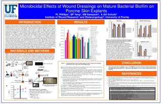
Microbicidal Effects of Wound Dressings on Mature Bacterial Biofilm on Porcine Skin Explants PL Phillips1, QP Yang1, EM Sampson2, & GS Schultz1 Institute of Wound Research1 and Otolaryngology2, University of Florida A B INTRODUCTION RESULTS A It has been hypothesized that four main causative factors, which can occur alone or in combinations, contribute to the pathogenesis of chronic wounds: tissue hypoxia, repetitive ischemia/reperfusion injury, age impaired stress response, and elevated bacterial levels. Persistent bacterial biofilm is known to contribute to molecular pathologies of many diseases, including cystic fibrosis, otitis media, and periodontitis.1 Recently, biofilms were proposed to be a major cause of impaired skin wound healing, contributing to the development of chronic wounds.2-4 James et al (2008) 4 showed that 60% of chronic wounds and 6% of acute wounds contained biofilm structures. Development of microbial biofilm, consisting of microorganisms embedded in a self-synthesized secreted exopolymeric matrix, forming complex dense microbial communities with complex 3D architecture provides substantial protection for bacteria to host antibodies, phagocytic inflammatory cells, antibiotics, antiseptics, and disinfectants while facilitating waste removal and uptake of nutritional requirements. Biofilm development is a complex process that is greatly influenced by the bacterial microflora, the environment, and in particular, the substrate to which it attaches. 5-7 We developed an in vitro model of mature biofilm cultured on porcine skin explants and used this model to assess the efficacy of commercial antimicrobial agents. Four types of antimicrobial agents (iodine, silver, polyhexamethylene biguanide (PHMB), and doxycycline) and three types of moisture dressings (cotton gauze, sodium carboxymethlcellulose fiber, and calcium alginate fiber), were assessed. Cadexomer iodine treatment produced complete kill of Pseudomonas aeruginosa (POA1) biofilm, whereas povidone iodine saturated gauze dressing reduced biofilm bacteria ~1-2 log. Nanocrystaline silver reduced PAO1 biofilm ~3 logs compared to its direct counterpart dressing. Dressings containing other forms of silver or PHMB did not significantly reduce biofilm bacteria. Doxycycline did not significantly reduce PAO1 biofilm levels compared to no dressing and ~1-2 logs compared to moisture dressings. Moisture dressings, particularly calcium alginate fiber, promoted PAO1 biofilm growth compared to no dressing. This model suggests that silver, povidone iodine, PHMB and doxycycline are relatively ineffective in killing mature Pseudomonas aeruginosa biofilm, whereas cadexomer iodine is an effective microbicidal wound dressing. *** Indicates a p<0.001 difference between each dressing and no dressing * * Indicates a p<0.05 difference between each dressing and no dressing *** *** *** ** ** ** *** *** *** Figure 3. Time course assessment of Iodine dressings on total Pseudomonas aeruginosa PAO1 bacteria (planktonic and biofilm) cultured on porcine explants. Freshly prepared sterile porcine explants inoculated with logarithmic planktonic bacterial suspension and cultured for 0, 1, 2, or 3 days was exposed to (A) Wet Gauze, (B) Povidone Iodine gauze, or (C) ddH2O hydrated Cadexomer Iodine dressing for 1, 12, or 24 hours. The explants were not treated with antibiotic for 24 hours to kill planktonic before dressing exposure. 0 day culture consists of explants inoculated with planktonic bacterial suspension, allowed to absorb for ~1 hour before addition of dressing. ● The results shows that Povidone iodine is effective in knocking down planktonic PAO1 bacteria indicated by the 0 and 1 day culture, but does not inhibit biofilm development nor kills PAO1 biofilm. Cadexomer iodine effectively penetrates the porcine explants and kill planktonic bacteria within 12 hours and killing PAO1 biofilm, knocking down biofilm bacteria below detectable levels (<100 CFU/ml) after 24 hours. C *** *** *** MATERIALS AND METHODS Figure 1. A) The key parameters of this model include preparing sterilized fresh porcine skin explants with partial thickness ‘wound beds’ inoculated with early logarithmic phase bacterial culture. The explants to be used in mature biofilm studies are treated for 24 hours in liquid media containing appropriate antibiotic to kill all planktonic bacteria to produce functionally mature biofilm. This study assessed the efficacy of commercially available antimicrobial and moisture wound dressings, after 1 or 3 days exposure, on mature 3 day Pseudomonas aeruginosa PAO1 biofilm cultured on porcine explants treated 24 hours with 200 μg/ml gentamicin (100 X MIC; Minimal inhibitory concentration). B). Growth curves show that PAO1 produce mature biofilm on porcine explants in after 3 days. *** *** CONCLUSION A 2 mm borehole • Figure 2. Antimicrobial Efficacy of Dressings on Mature 3 Day Pseudomonas aeruginosa PA01 Biofilms after 1 and 3 Days of Dressing Exposure • Iodine is the most effective antimicrobial agent tested against mature 3 day Pseudomonas aeruginosa PAO1 biofilm and Cadexamer Iodine is the most effective form. • Cadexomer iodine (Iodoflex™) reduced mature PAO1 biofilm ~8 logs (from ~108 to <100 CFU/ml, the detection limit of the spread plate quantification method used) after 1 and 3 days exposure to dressing completely hydrated with sterile ddH2O. Overnight TSB culture of 1 and 3 day exposed mature PAO1 biofilm explants had no growth verifying complete biofilm kill. • Povidone iodine saturated Gauze reduced mature PAO1 biofilm ~1.5-2 logs after 1 day exposure compared to no dressing or wet gauze sponge. • Silver only partially kills mature PAO1 biofilms. • The results showing that Acticoat™–Absorbant significantly reduces biofilm compared to it’s direct counterpart dressing Algisite™- M by ~3 logs, suggesting that nanocrystaline silver is the most effective form of antimicrobial silver. • This antimicrobial effect was not observed when comparing Aquacel-Ag/Aquacel, Acticoat ™ -7/Gauze, or Tegaderm-Ag/Gauze dressing pairs. • 3% Doxycycline (NanoDox™ hydrogel) does not significantly reduce PAO1 biofilm compared to no dressing and ~1 - 1.5 log compared to moisture dressings. • 0.2% polyhexamethylenebiguanide (PHMB) (Curity™ AMD™) does not significantly reduce PAO1 biofilm compared to no dressing or moisture dressings. • Moisture dressing, particularly calcium alginate (Algisite™-M), appears to promote biofilm growth to a small extent. • Due to less available moisture and nutrients (the dressings were hydrated and absorbed nutrients and antibiotic from the media), the antimicrobial dressings are most appropriately compared to their corresponding dressing lacking the antimicrobial rather than no dressing. Prepare 8mm Explants Colony count (CFU / ml) Chlorine Gas Explant Sterilization Plate 100 µl in triplicate This model showed that antibiotics, silver, and PHMB are the least effective in killing existing mature Pseudomonas aeruginosa biofilm. Cadexomer iodine appears to be an effective antimicrobial wound dressing. Innoculate Explants 10-1 - 10-6 Dilution factor REFERENCES 150 µL 10 µL 5ml TSB Serial dilute 37°C overnight Soft 0.5% agar TSA + antibiotics OD640 0.2-0.4 Sonicate in 7 mL PBS with 5 µl/ml Tween 80 Bacteria agar streak plate 50 mL TSB Donlan RM, Costerton JW. Biofilms: survival mechanisms of clinically relevant microorganisms. Clin Microbiol Rev 2002; 15: 167-193. Jones SG, Edwards R, Thomas DW. Inflammation and Wound Healing: the Role of Bacteria in the Immuno-Regulation of Wound Healing. Int J Low Extrem Wounds 2004;3:201-8. Edwards R, Harding KG. Bacteria and Wound Healing. Curr Opin Infect Dis 2004;17:91-6. James GA, Swogger E, Wolcott R, Pulcini E, Secor P, Sestrich J, Costerton JW, Stewart PS. Biofilms in Chronic Wounds. Wound Repair Regen 2008;16:37-44. Ren D, Bedzyk LA, Thomas SM, Ye RW, Wood TK. Gene Expression in Escherichia Coli Biofilms. Appl Microbiol Biotechnol 2004;64:515-24. Cho KH, Caparon MG. Patterns of Virulence Gene Expression Differ Between Biofilm and Tissue Communities of Streptococcus Pyogenes. Mol Microbiol 2005;57:1545-56. Luppens SB, ten Cate JM. Effect of Biofilm Model, Mode of Growth, and Strain on Streptococcus mutans Protein Expression As Determined by Two-Dimensional Difference Gel Electrophoresis. J Proteome Res 2005;4:232-7. 24 hr TSB + Antibiotic Culture 1-5 days 37°C 5% CO2 Transfer to fresh plates daily Exposure to ddH2O hydrated dressing Weighted with sterile slide B PAO1 Growth Curve of 1-5 day biofilm Mature PAO1 formed on explants after 3 days. Bacterial levels are expressed as CFU /mL found in the 7 mL PBS/Tween 80 sonicated bacterial suspension.


