Bowel obstruction & Hernias
Bowel obstruction & Hernias. Hugh Tulloch. Learning objectives. Go through the basics of hernias and bowel obstruction Anatomy Dapsicamp Focus on inguinal and femoral hernias . What is the definition of a hernia??. Hernias. Definition:


Bowel obstruction & Hernias
E N D
Presentation Transcript
Bowel obstruction&Hernias Hugh Tulloch
Learning objectives • Go through the basics of hernias and bowel obstruction • Anatomy • Dapsicamp • Focus on inguinal and femoral hernias
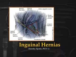
Hernias • Definition: • A protrusion of a viscus (organ) or part of a viscus through a defect in a wall that contains it.
epidemiology • Comprise of 7% of all surgical outpatients • 1-3% of young children (congenital) • Most common in elderly men (200/10000 person years) – 75 years
Question time • Names some common hernias
Common hernias • Inguinal, femoral, umbilical, incisional • Can further be described: • Reducible • Irreducible (incarcerated) • Strangulated • Richter’s – only lumen wall is herniated (strangulation without obstruction)
Inguinal hernias • Male > female • Indirect (80%) • Follows normal course of inguinal canal • May have congenital origin – failure of regression of processusvaginalis • Direct (20%) • Does not go through deep ring. • Weakness in abdo wall • **Above and medial to pubic tubercle**
Background - anatomy • Inguinal ligament
Inguinal canal • The anterior wall is formed by the aponeurosis of the external oblique, and reinforced by the internal oblique muscle laterally. • The posterior wall is formed by the transversalis fascia. • The roof is formed by the transversalis fascia, internal oblique and transversusabdominis. • The floor is formed by the inguinal ligament (a ‘rolled up’ portion of the external oblique aponeurosis) and thickened medially by the lacunar ligament.
The two openings to the inguinal canal are known as rings. The deep (internal) ring is found above the midpoint of the inguinal ligament. which is lateral to the epigastric vessels. The ring is created by the transversalisfascia, which invaginates to form a covering of the contents of the inguinal canal. • The superficial (external) ring marks the end of the inguinal canal, and lies just superior to the pubic tubercle. It is a triangle shaped opening, formed by the evagination of the external oblique
Contents of inguinal canal3: arteries, nerves, fascial layers, others • The structures which pass through the canal differ between males and females: • in males : the spermatic cordand its coverings + the ilioinguinal nerve. • in females : the round ligament of the uterus + the ilioinguinal nerve.
Spermatic cord • 3 arteries: artery to vas deferens (or ductus deferens), testicular artery, cremasteric artery; • 3 fascial layers: external spermatic, cremasteric, and internal spermatic fascia; • 3 other structures: pampiniform plexus, vas deferens (ductus deferens), testicular lymphatics; • 3 nerves: genital branch of the genitofemoral nerve (L1/2), sympathetic and visceral afferent fibres, ilioinguinal nerve (N.B. outside spermatic cord but travels next to it) • Note that the ilioinguinal nerve passes through the superficial ring to descend into the scrotum, but does not formally run through the canal.
Femoral • Female > male • Increased risk of strangulation • **Lateral and inferior to pubic tubercle** • Femoral canal: • • Boundaries: • ANTERIOR: inguinal ligament • POSTERIOR: pectineal ligament • MEDIAL: lacunar ligament • LATERAL: femoral vein
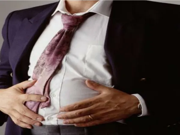
History • Onset, course, duration • Painful? (Socrates) • Other, previous lumps • Symptoms of bowel obstruction • Possible causes: COPD, cough, urinary obstruction • Increase abdo pressure • Does lump disappear? -reducible
Differentials • Other hernias – femoral, inguinal etc • Enlarged lymph nodes • Ectopic testis • Femoral aneurysm
Bowel obstruction • Aetiology Can be caused by a physical blockage or due to a ‘shocked’ bowel • Mechanical • Physical object blocking • Lumenal, extra luminal • Non-mechanical • Surgical • Postopertaive, peritonitis, ischaemia • Medical • Electrolyte abnormalities • Drugs – opiates, anticholinergics
Gallstone ileus -cholecysto-enteric fistula ”an abnormal connection between an organ, vessel, or intestine and another structure”
In general… • Small bowel • Adhesions and hernias • Large bowel • neoplasia, diverticulitis, volvulus
Non mechanical obstruction • Ileus • disruption of the normal propulsive ability of the gastrointestinal tract. • Paralytic ileus • postsurgical ileus – gastrointestinal surgery • Electrolyte imbalance • Hypothyroidism • Spinal cord injury
Question time • What would an obstructed patient complain of?
Clinical features - symptoms • 4 cardinal symptoms • 1. pain • Small bowel – central • Large bowel – lower and colicky (if constant worry about perforation) • 2. constipation (absolute) • 3. vomiting • 4. distention
investigations • Imaging • Erect CR and AXR • U&Es – electrolyte abnormalities
Question time • Why would you perform an erect CXR?
Differences… (first) (second)
Treatment - mechanical • Mostly conservative • Nil by mouth and NG tube • Analgesia • Surgery indications: • Strangulation or perforation • Closed loop obstruction • Failure of conservative approach • neoplasms
Non mechanical • Conservative • -prevention (reduce bowel handling in surgery) • Analgesia • NBM and NG tube • Medical – electrolyte restoration
prognosis • Depends on type.. • Non-complicated have mortality 3-5% • If ischaemic can be 30%
Question time • What features on a AXR would indicate small bowel obstruction? • Fluid levels, central position of loops, valvulaeconniventes (all the way across) • Where would you see a femoral hernia • Below and lateral • What is the definition of a hernia? • A protrusion of a viscus (organ) or part of a viscus through a defect in a wall that contains it.
Case study • Mr X comes in with intramittent central abdo pain. He is constipated, vomiting. On examination his stomach is distended. You also notice a lump in his groin • What is your differential • Inflamatory – appendicitis? Perforation of ulcer? • Vascular - occlusive intestinal ischemia, usually caused by thromboembolism of the superior mesenteric artery • What investigations do you order • CXR, AXR • What more information about the lump would you want to know • Reducible? Location? Painful?
Case study • Mrs Y has just undergone surgery to remove a colorectal cancer • Her chart reads BNO for the last 3 days • What does BNO stand for? • What do you think the cause is? • How should you manage her?