Download
1 / 30
300 likes | 317 Vues
The incidence of chronic kidney disease CKD and end stage renal disease ESRD has been increasing steadily for decades. The CKD epidemic is a worldwide public health problem, associated with premature death, increased morbidity and, last but not least, enormous costs related to renal replacement therapy RRT . The rise in ESRD globally is largely due to the increasing prevalence of diabetes mellitus and nephrosclerosis, the two major causes of CKD An increasing number of older patients, particularly those over 65 years of age, is also a contributing factor. CKD is usually associated with a progressive loss of renal function over several years or even decades. When patients with CKD progress to Stage 5 renal failure glomerular filtration rate GFR below 15ml min 1.73m2 they usually suffer from uraemic symptoms at this stage the possibility of starting RRT should have already been discussed. There are two options for RRT chronic dialysis therapy peritoneal dialysis or haemodialysis or renal transplantation RTx . The present work relates primarily to kidney transplant patients. Dr. Amit Kumar Verma "Bony Changes in CKD Patient with or Without Hemodialysis" Published in International Journal of Trend in Scientific Research and Development (ijtsrd), ISSN: 2456-6470, Volume-2 | Issue-6 , October 2018, URL: https://www.ijtsrd.com/papers/ijtsrd18567.pdf Paper URL: http://www.ijtsrd.com/medicine/other/18567/bony-changes-in-ckd-patient-with-or-without-hemodialysis/dr-amit-kumar-verma<br>

E N D
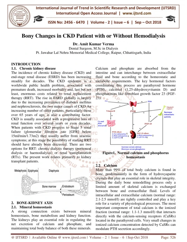
International Journal of Trend in Scientific Research and Development (IJTSRD) International Open Access Journal | www.ijtsrd.com ISSN No: 2456 - 6470 | Volume - 2 | Issue – 6 | Sep – Oct 2018 Bony Changes in CKD Patient with or Without Hemodialysis Dr. Amit Kumar Verma Dental Surgeon, M.Sc in Dialysis Pt. Jawahar Lal Nehru Memorial Medical College, Raipur, Chhattisgarh,India INTRODUCTION 1.1. Chronic kidney disease The incidence of chronic kidney disease (CKD) and end-stage renal disease (ESRD) has been increasing steadily for decades. The CKD epidemic is a worldwide public health problem, associated with premature death, increased morbidity and, last but not least, enormous costs related to renal replacement therapy (RRT). The rise in ESRD globally is largely due to the increasing prevalence of diabetes mellitus and nephrosclerosis, the two major causes of CKD An increasing number of older patients, particularly those over 65 years of age, is also a contributing factor. CKD is usually associated with a progressive loss of renal function over several years or even decades. When patients with CKD progress to Stage 5 renal failure (glomerular filtration rate [GFR] below 15ml/min/1.73m2) they usually suffer from uraemic symptoms; at this stage the possibility of starting RRT should have already been discussed. There are two options for RRT: chronic dialysis therapy (peritoneal dialysis or haemodialysis) or renal transplantation (RTx). The present work relates primarily to kidney transplant patients. Calcium and phosphate are absorbed from the intestine and can interchange between extracellular fluid and bone according to the homeostatic and metabolic requirements. The key hormonal factors coordinating this process are parathyroid hormone (PTH), calcitriol (1,25-dihydroxyvitamin D) and phosphatonins like fibroblast growth factor 23 (FGF- 23). Figure1. Normal calcium and phosphorus homeostasis 2.2. Calcium More than 99% of total body calcium is found in bone, predominantly in the form of hydroxyapatite crystals that play an essential role in skeletal integrity. During the daily bone remodelling process only a limited amount of skeletal calcium is exchanged between bone and extracellular fluid. Levels of intracellular and extracellular calcium (normal range: 2.1-2.5 mmol/l) are tightly controlled and play a key role for a variety of physiological processes. The most important component of total calcium is the ionized fraction (normal range: 1.1-1.3 mmol/l) that interacts directly with the calcium-sensing receptors (CaSRs) located in the parathyroid cells. Very small changes in ionized calcium concentration detected by CaSRs can modulate PTH secretion accordingly. 2. BONE-KIDNEY AXIS 2.1. Mineral homeostasis A strong connection exists between mineral homeostasis, bone metabolism and kidney function. The kidneys play an essential role in regulating the net excretion of calcium and phosphate, and maintaining total body balance of both these minerals. @ IJTSRD | Available Online @ www.ijtsrd.com | Volume – 2 | Issue – 6 | Sep-Oct 2018 Page: 526
International Journal of Trend in Scientific Research and Development (IJTSRD) ISSN: 2456-6470 2.3. Phosphate Phosphate is necessary for matrix mineralization and most of it (85%) is present in bone, with only 0.1% located in the extracellular space. Phosphate is present in cell membranes (phospholipids), and nucleic acid (RNA, DNA), and participates in energy metabolism of all biological systems. The maintenance of phosphate homeostasis, therefore, is of crucial biological importance. About 90% of inorganic plasma phosphate (Pi) is ultra-filterable, emphasizing the role of the kidney in phosphate excretion. Normal plasma concentration is maintained between 0.7-1.5 mmol/l. Until recently, phosphate homeostasis had been thought to be regulated by the same factors involved in maintaining calcium homeostasis i.e. PTH, calcitonin and vitamin D (1, 25(OH) 2D3). Recently, however, another hormone, FGF-23, has been identified that principally regulates phosphate homeostasis and bone formation. 2.4. Hormonal factors of mineral homeostasis 2.4.1.Parathyroid hormone Biologically active PTH (1-84) is synthesized in parathyroid cells and released timely to maintain serum calcium concentrations within a tight physiologic range. When the serum calcium concentration drops, CaSRs become activated in the parathyroid cells and facilitate PTH release. PTH enhances the active reabsorption of calcium in the distal tubules and increases the absorption of calcium from the bowel by stimulating the synthesis of 1, 25(OH) 2D3. Furthermore, PTH indirectly affects bone degradation by stimulating the binding of the receptor activator of nuclear factor kappa-B ligand (RANKL) to RANK, osteoclastogenesis followed by bone resorption. PTH undergoes metabolic degradation minutes. Intact PTH (iPTH; 1- 84) and a fragment with an intact N-terminus, PTH (1-34), have the greatest biological activity. In the general population, the average PTH level is 1.1-6.9 pmol/l. As PTH is eliminated via glomerular filtration and tubular degradation, these fragments can accumulate in kidney failure 2.4.2. Calcitonin Calcitonin is peptide hormone secreted by specialized cells called C-cells in the thyroid gland. An increase in serum calcium stimulates calcitonin secretion. It decreases bone resorption by inhibiting osteoclasts through direct action on the osteoclastic calcitonin receptors. 2.4.3.Fibroblast growth factor 23 –“a new player in the field” FGF-23 is a circulating phosphaturic hormone that plays an important role in phosphate homeostasis by regulating renal phosphate excretion via inhibition of Na-Pi-2a co-transport in the kidney. It is principally produced by osteocytes in response to increased serum phosphate. FGF-23 receptors are located in proximal tubular cells. Interaction between these receptors and a transmembrane protein called Klotho leads to the inhibition of phosphorus reabsorption and to impaired 1,25(OH)2D3 production via the inhibition of 1-α-hydroxylase . Recently, a vitamin D response element site was identified in the FGF-23 promoter region and it appears that 1, 25(OH) 2D3 stimulates FGF-23 activity in osteocytes. Therefore, FGF-23 can be considered as a counter- regulatory hormone for 1,25(OH)2D3 that maintains phosphate balance . Its role is summarized in Figure 2. FGF-23 levels increase as renal function declines in response to phosphate retention. Depending on the assay used, the normal range of FGF-23 is approximately10 to108 RU/ml. resulting increased within Figure 2; A simplified diagram of the regulation of calcium and phosphorous balance @ IJTSRD | Available Online @ www.ijtsrd.com | Volume – 2 | Issue – 6 | Sep-Oct 2018 Page: 527
International Journal of Trend in Scientific Research and Development (IJTSRD) ISSN: 2456-6470 Figure 3; the regulation and action of FGF-23 2.4.4. Vitamin D Precursors for active vitamin D hormones derive from dietary intake (plant sources, ergocalciferol - vitamin D2, and from animal sources cholecalciferol - vitamin D3) or from 7-dehydrocholesterol in the skin during sun exposure. Two important hydroxylation steps are necessary in the metabolism of vitamin D in order for its hormonal active forms to be produced. The first step is relatively fast and unregulated, and requires 25-hydroxylase enzymes in the liver to produce 25- hydroxyvitamin D (25- OHD). The biological half-life of serum 25-OHD is approximately 2-3 weeks, whereas the half-life of the active hormone, 1,25(OH)2D3 is only 4-6 hours. Measuring plasma 25-OHD level represents the best index of nutrition vitamin D intake. Further hydroxylation occurs by renal 1-α-hydroxylase located in cells of the proximal tubule, and results in the formation of the active hormone, calcitriol. Calcitriol enters the target cell and binds to the vitamin D receptor (VDR). The liganded VDR then translocates to the nucleus where it interacts with the target DNAs and regulates gene transcription. Calcitriol is the major regulator of active intestinal calcium absorption and has a major effect on the differentiation of osteoclasts via the calcitriol- induced binding of RANKL to the RANK receptor . The renal synthesis of calcitriol is regulated by calcium, phosphorus, FGF-23 and PTH. Low levels of calcium or phosphorus and increasing PTH level promote calcitriol synthesis. described above, FGF-23 inhibits the activity of 1-α- hydroxyalse.The normal concentration in healthy individuals is 20-60 pg/ml. This decreases progressively as kidney function declines. According to the Kidney Disease Outcomes Quality Initiative (K/DOQI) guidelines, patients with CKD should undergo biochemical screening to detect vitamin D deficiency and receive timely treatment if needed. Plasma 25-OHD levels between 16 and 30 ng/ml qualify as vitamin D insufficiency, and values ranging from 5 to 15 ng/ml indicate vitamin D deficiency. These conditions can be treated with ergocalciferol, which should not be mistaken for the active vitamin D hormone (calcitriol) that is used to treat secondary hyperparathyroidism. Both inadequate vitamin D (25-OHD) and impaired calcitriol (1,25(OH)2D3 ) production can be observed in patients with CKD. 3. BONE PHYSIOLOGY 3.1. Bone structure Bone is a complex, highly organized and specialized connective tissue. Its organic is composed of collagen fibers, which provide flexibility. The hard matrix of calcium salts (hydroxyapatite) deposited around the collagen fibers makes the bone rigid. There are two major types of bone in the human skeleton: cortical and trabecular. Nearly 80% of the total skeletal mass is cortical bone providing structural strength, while the rest is trabecular. The proportion of trabecular and cortical bone varies at different skeletal regions. In the vertebrae and the ultradistal forearm, trabecular bone makes up approximately 70% of the bone. In contrast, the proximal third of the radius consists entirely of cortical bone and the femur neck approximately 75%. The outer surface of bone is covered by periosteum with the endosteum lining the internal surface. There are two major categories of bone cell: osteoclast and osteoblast Furthermore, as serum 1,25(OH)2D3 @ IJTSRD | Available Online @ www.ijtsrd.com | Volume – 2 | Issue – 6 | Sep-Oct 2018 Page: 528
International Journal of Trend in Scientific Research and Development (IJTSRD) ISSN: 2456-6470 3.2. Bone remodelling Bones are constantly undergoing remodelling via bone resorption by osteoclasts and bone formation by osteoblasts. Trabecular bone, with its large surface, is the predominant site of bone remodelling. Several hormones and local factors are involved in the control of this process. In normal adults, bone resorption and bone formation are well balanced. Imbalance between the two processes – i.e. osteoclast activity exceeding 18 osteoblast activity – leads to decreased bone mass and an increased risk of osteoporotic bone fracture. 3.3. Bone disease Mineral and bone disorders in chronic kidney disease (CKD-MBD) As CKD progresses, various bone disorders may develop. The classical term “renal osteodystrophy” has recently been replacedby “CKD- Mineral and bone disorder (CKD- MBD)” and is characterized by three components: laboratory abnormalities (of calcium or phosphorus or PTH or vitamin D), bone disease, and vascular calcification [20]. The classification of renal bone diseases is based on histological findings. High and low turnover bone diseases have been categorized based on over- stimulated or over- suppressed PTH, respectively. High-turnover bone diseases, characterized by increased bone remodelling, include osteitis fibrosa caused by secondary hyperparathyroidism (SHPT) and mixed disorders. The vast majority of CKD patients have some degree of SHPT [10, 20, 21]. The pathophysiology of SHPT is illustrated in Figure . Figure4. Calcium and phosphorus metabolism in renal failure Low-turnover bone disease includes osteomalacia and adynamic bone disorder (ABD). Osteomalacia develops because of mineralization. This was mainly caused by aluminum- based medical interventions which have now generally been abandoned so osteomalacia has become a rare condition. ABD is characterized by a low number of osteoblasts with decreased bone formation caused by inappropriate suppression of PTH inappropriate bone Table1. Effect of immunosuppressive treatment on bone Immunosuppressive agent Effect Reduce intestinal calcium absorption Increase urinary calcium excretion Decrease the effect of vitamin D Increase parathyroid hormone Decrease adrenal and gonadal steroid synthesis Decrease osteoblastic bone formation Increase osteoclasts and bone resorption Resorption rate higher than bone formation Resorption higher than formation (probably less severe than CsA) No effect on bone No effect on bone Hypophosphataemic osteomalacia by increasing hyperphosphaturia Inhibits longitudinal bone growth Glucocorticoids Systemic effects Local effect on bone Calcineurin inhibitors Cyclosporine A (CsA) Tacrolimus (FK506) Azathioprine Mycophenolate mofetil mTOR inhibitor @ IJTSRD | Available Online @ www.ijtsrd.com | Volume – 2 | Issue – 6 | Sep-Oct 2018 Page: 529
International Journal of Trend in Scientific Research and Development (IJTSRD) ISSN: 2456-6470 4. DIAGNOSIS DISEASE 4.1. Bone biopsy The most accurate diagnostic test for determining the type of renal osteodystrophy is transiliac bone biopsy with double tetracycline histomorphometric analysis. Bone biopsy is the gold standard, but as it is an invasive and painful procedure, it is infrequently used in routine clinical practice. It also requires technical expertise and a well-trained histomorphometrist to analyze the samples. At present, bone biopsy analysis by dynamic histomorphometry is not performed in Norway 5. MEASUREMENT OF BONE MINERAL DENSITY 5.1. Dual-energy x-ray absorptiometry Dual-energy x-ray absorptiometry (DXA) is a widely used, non-invasive method measuring bone mineral content (g/cm2) osteopenia/osteoporosis. The technique implies a short scanning time (about 10 minutes), low radiation exposure (approximately < 1μSv), low precision error OF METABOLIC BONE independent of the operator (0.5-2%), and acceptable costs. The preferred skeletal sites for BMD measurement using DXA are the lumbar spine, total femur, radius, and whole body. The results of BMD measurements can be expressed as absolute values (g/cm2), or as T- score and Z-score T-score: the number of standard deviations above or below the mean reference value for a healthy 30 year old adult of the same sex as the patient. Z-score: the number of standard deviations above or below the mean reference value for the patient's age and sex. Patients can be classified according to the World Health Organization (WHO) osteoporosis (Table 2) [52]. However, it is important to note that white, post- menopausal population was used to define the WHO criteria. This has limited applicability to patients immunosuppressants, or more heterogeneous patient populations (e.g. males, mixed ethnicity). labeling and bone definition of and identifying treated with Table2. World Health Organization definition of osteoporosis Fracture Risk Very low 4x 8x Classification Definition Normal Osteopenia Osteoporosis Severe osteoporosis BMD no more than 1 SD below the young adult mean BMD is 1 to 2.5 SD below the young adult mean (T score –1 to –2.5) BMD >2.5 SD below the young adult mean (T score >–2.5) BMD >2.5 SD below the young adult mean plus history of one or more fragility fractures 20x Bone loss is more prevalent in trabecular bone, however, cortical bone, characterized by high bone mineral content, can hide small changes in trabecular bone during the DXA assessment. As DXA does not provide specific information on bone turnover and the method is unable to differentiate between trabecular and cortical bone, the BMD results should be interpreted together with clinical assessment, bone biomarkers and bone histology if it is possible. 5.2. Quantitative computed tomography Another non-invasive procedure is quantitative computed tomography (QCT) that we did not use in our study. It measures BMD accurately and provides a three-dimensional image with separate assessment of cortical and trabecular bone. This technique measures the volumetric size (mg/ml) of the target bone. The main disadvantage of QCT is the high radiation exposure and a high cost. 5.3. Biochemical markers of bone metabolism While bone histology is still the gold standard for the accurate assessment of bone turnover, the search for reliable biochemical bone markers has been ongoing for several years. With the development of new, more sensitive and specific assays, our ability to determine bone turnover by biochemical markers has improved considerably. Though measurement of iPTH has been still widely used and it provides basis for the assessment of bone turnover, serum iPTH levels alone are insufficient to clearly distinguish adynamic or normal bone from hyperparathyroid bone disease. Therefore the specificity of PTH as an indicator of bone turnover has been questioned. According to Qi et al., serum iPTH levels between 65 and 450 pg/ml could not predict the degree of bone turnover in dialyzed patients, and bone biopsy was proposed to these patients. A number of biochemical markers are available clinically to assess bone metabolism in @ IJTSRD | Available Online @ www.ijtsrd.com | Volume – 2 | Issue – 6 | Sep-Oct 2018 Page: 530
International Journal of Trend in Scientific Research and Development (IJTSRD) ISSN: 2456-6470 patients with chronic renal failure. Biochemical markers respond within days or weeks after initiation of anti-resorptive therapy. Biochemical markers of bone resorption respond considerably faster than the markers of bone formation. All biomarkers have limitations and their clinical applicability remains to be established. Panels of simultaneously obtained bone formation and bone resorption markers need to be analyzed for an appropriate evaluation of bone disease in routine clinical practice. The most appropriate application for these biomarkers is to monitor trends over time. 6. MARKERS OF BONE FORMATION Osteocalcin: Osteocalcin is a major non-collagenous protein of the bone matrix produced by osteoblasts. Osteocalcin is released into the bloodstream during bone matrix synthesis. At the ‘local’ bone level, osteocalcin plays an important role in bone mineralization and may also induce bone resorption by stimulating adhesion and chemotaxis of osteoclasts . At the ‘systemic’ level, recent data have shown that osteocalcin regulates pancreatic β cell proliferation and adipocyte gene expression, increasing insulin secretion and adiponectin synthesis, respectively. Its physiological role in bone energy regulation is shown in Figure 4 . Osteocalcin has a short half-life in the blood and its serum concentration is determined mainly by renal clearance. In patients with CKD, serum osteocalcin increases as renal function declines. Figure5. Hypothetical relationships between osteocalcin and adiponectin Bone-specific alkaline phosphatase: Alkaline phosphatase (ALP) is a cell- membrane- associated enzyme expressed mainly by the liver and bone in adults. In bone, ALP is derived from osteoblasts and it influences bone mineralization. In the absence of liver failure, ALP is a useful indicator of osteoblast activity. Furthermore, bone-specific alkaline phosphatase (BSAP) is the fraction of total alkaline phosphatase that is specific to the osteoblast. The serum level of BSAP is not influenced by renal failure. There is evidence that BSAP correlates well with iPTH levels and histomorphometric indices of SHPT in CKD patients. Propeptides of collagen type I: Type I collagen is synthesized by osteoblasts from type I procollagen precursor proteins (Figure 5). These precursors have large extension domains at both ends. When type I collagen is synthesized, these propeptides, procollagen type 1 carboxy-terminal extension peptids (PINP) and procollagen type 1 amino-terminal extension peptids (PICP) respectively, are enzymatically removed and released into the circulation. Higher levels of PINP and PINP in the plasma may indicate increased bone formation. PINP and PICP are degraded in the liver. @ IJTSRD | Available Online @ www.ijtsrd.com | Volume – 2 | Issue – 6 | Sep-Oct 2018 Page: 531
International Journal of Trend in Scientific Research and Development (IJTSRD) ISSN: 2456-6470 Figure 6 Schematic representation of a type I pro-collagen molecule 7.MARKERS OF BONE RESORPTION Collagen breakdown products: The organic matrix of bone consists of type I collagens, which are held together by pyridinoline and deoxypyridinoline cross- links and provide mechanical force to the bone tissue. When collagen degrades, these cross-links and other fragments are released into the circulation, and excreted in the urine. The breakdown of type I collagen is mediated by acid proteases derived from osteoclasts. Telopeptides are small amino acid sequences originating from the nonhelical ends of collagen molecules as a result of enzymatic degradation. Fragments released by this process include N-, and C-terminal cross-linked telopeptides (NTX, CTX), procollagen type I cross-linked carboxy-terminal telopeptide (ICTP) and pyridinoline (PYD) and deoxypyridinoline (DPD) . We studied only the role of CTX in bone turnover Tartrate- resistant acid phosphatase (TRAP): Tartrate-resistant acid phosphatase is produced by osteoclast during bone resorption, and the TRAP 5b isoform has recently been identified as the osteoclast-specific portion of TRAP. During bone degradation, TRAP5b is released into the circulation and is cleared mainly by the liver. The TRAP 5a isoform is a marker of inflammatory conditions and is synthesized by macrophages. We have not measured its level in our study. A.K/DOQI - General recommendations a.Use the lowest possible steroid dose b.Stop smoking c.Initiate exercise d.Treat persistent hyperparathyroidism ➢Optimal management of calcium ➢Optimal management of vitamin D ➢Possible role of calcimimetics ➢Parathyroidectomy e.Avoid loop diuretics if possible f.Substitute for insufficient gonadal or thyroid function B.Calcium and vitamin D supplements Vitamin D metabolism is disrupted before and after kidney transplantation. It is recommended that transplant recipients with good kidney function and normal serum calcium level receive calcium (1000 to 1500 mg/day) and vitamin D in the form of cholecalciferol or ergocalciferol (400 to 800 IU/day) supplements in order to moderate post-transplant bone loss . Small doses (0.25-0.5 dg/day) of active vitamin D derivates – alfacalcidiol, doxercalciferol or calcitriol have also had a positive effect on BMD at the lumbar spine and femoral neck in kidney transplant patients. 8. BONE MINERAL DISORDERS IN CKD Chronic kidney disease (CKD) is associated with numerous metabolic and nutritional alterations affecting among others mineral metabolism and bone health which are interrelated and together form an entity called CKD-mineral and bone disorders (CKDMBD). CKD-MBD disturbances of phosphate and calcium homeostasis as well as with changes in key regulators of bone status such as parathyroid hormone (PTH) and fibroblast growth factor 23 (FGF23). These alterations may lead to renal osteodystrophy with bone loss, osteoporosis, and potentially fractures, and increased risk for premature vascular calcification, adding significantly to other common causes of cardiovascular disease (CVD), the leading cause of death in CKD patients. CKD-MBD is thus thought to be a major contributor to the high mortality among patients with CKD. is associated with @ IJTSRD | Available Online @ www.ijtsrd.com | Volume – 2 | Issue – 6 | Sep-Oct 2018 Page: 532
International Journal of Trend in Scientific Research and Development (IJTSRD) ISSN: 2456-6470 While many of the circulating mediators of CKD- MBD can be relatively easily measured, a detailed assessment of bone status requires more complex methods some of which such as bone biopsy are not readily available. However, the measurement of bone mineral density (BMD) usually performed by dual- energy X-ray absorptiometry (DEXA) is a more convenient method in the clinical setting and is increasingly regarded as an integral component of assessment of bone mass, presence and extent of osteoporosis, and risk of fractures. BMD is a well- established key parameter for monitoring bone disease in CKD patients, although it should be noted that, there are many common factors affecting BMD not specifically related to CKD-MBD, such as age, gender, menopause, estrogen consumption, body mass, cigarette smoking, alcohol abuse, excess glucocorticoid exposure, physical activity and genetic factors . It should also be noted that BMD cannot fully describe the status of bone fragility since bone status is due to many dynamic factors such as abnormal bone turnover and remodeling, leading to impairment of bone micro-architecture. higher but variable depending both on population- specific characteristics and on the techniques and body sites used for measurements. The effects of renal replacement therapy on bone mass vary between the different therapies. Although renal transplantation results in improvement of many aspects of CKD-MBD, it may further worsen bone mineral deficiency. BMD measured in the lumbar bone decreased significantly after six months of transplantation, which seemed to be mediated primarily by glucocorticoid transplantation. usage after On the other hand, it was reported that in patients on dialysis treatment, BMD did not change during the first year of dialysis treatment, neither in patients on hemodialysis (HD) nor in patients treated by peritoneal dialysis (PD). Whereas an increased risk of hip fracture was found to be related to the duration of dialysis, the impact of dialysis treatment as such on BMD is still not clear. There is a positive correlation between body weight and BMD in the general population. Similarly, several studies demonstrated that the body size relates with BMD also in CKD and ESRD patients. In general body mass index (BMI) associates with BMD or bone mass as measured by DEXA and BMI is a predictor of BMD also in ESRD patients. Thus, not surprisingly, body weight and BMI were primary responsible factors for BMD variation, osteoporosis and fractures, and were determinants also of the Z-score of BMD at mid-radius, femoral neck, lumbar spine and measurement sites, and total body BMD, in HD patients. It should be noted however that the relationship between BMI and BMD in the lumbar spine area is confounded by presence of aortic calcification leading measurements of BMD. Obesity also is associated with elevated BMD of weight-bearing bones and ribs, suggesting that obesity per se or indirectly via the increase of body mass may prevent bone loss. On the other hand, reduced lean body mass and sarcopenia associated with bone loss as demonstrated in in elderly people, putatively due to reduced impact on bone remodeling which through increased mechanical load forces of lean tissue may serve to strengthen bone. It is not surprising that a large body mass is closely related with high BMD also in CKD patients and body mass was reported to correlate with BMD at all body sites in HD patients. Disruption in mineral metabolism occur already at early stages of CKD, leading as the disease progresses to alterations in bone mass, bone turnover, mineralization and bone health . Disorders of bone structure and bone mass may result in severe osteoporosis and marked risk of fractures. Moreover, and even more important, because of the close links between bone status and soft tissue calcification, these alterations associate with vascular calcification, sometimes described as vascular ossification, leading to clinically manifest CVD and increased mortality. The following brief review which in part is based on a recent review article from our group summarizes the current understanding of causes of CKD-MBD, and its consequences for clinical outcome in CKD patients. to erroneously high 9. CAUSES OF IMPAIRED BMD IN CKD 9.1 Renal function, body weight and age BMD decreases progressively in patients during the course of progression from mild to severe degrees of renal failure and the decrease in BMD is thus most pronounced in patients glomerularfilltration rate (GFR). Bianchi et al. reported that patients with pre-dialysis renal failure have reduced BMD which correlated to the reduction of renal function. In end-stage renal disease (ESRD), the reported prevalence of bone mineral deficiency is with the lowest @ IJTSRD | Available Online @ www.ijtsrd.com | Volume – 2 | Issue – 6 | Sep-Oct 2018 Page: 533
International Journal of Trend in Scientific Research and Development (IJTSRD) ISSN: 2456-6470 The association of body fat mass with bone status may in addition be related to the metabolic activity of adipose tissue potentially affecting the skeleton as well as by hormonal alterations linked at least in part to the amount of adipose tissue, such as alterations in vitamin D metabolism, production, free fraction of sex steroids, insulin levels, and leptin levels. Thus, a high calcium dialysis solution may protect against osteoporosis whereas a low calcium dialysate may increase the risk of osteoporosis and hyperparathyroidism especially during long-term use . Some studies found an inverse correlation between PTH levels and mid-radius BMD reected by Z-scores in HD patients. Others could not find such correlation of PTH and BMD in dialysis patients. These discrepancies may be due to differences in follow-up time. While BMD mainly reflects long-term influence of CKD-MBD factors, PTH has more immediate effects on the metabolic state of bone in CKD patients and an increased PTH level will probably not result in substantial bone loss in the short-term. Another factor of importance is vitamin D which was reported to be linked to BMD, but not to fracture incidence, in patients with CKD stage 3 to 5 patients. Vitamin D supplementation reduces serum PTH levels and improves bone strength in animal studies. peripheral estrogen Appropriate regular physical activity and especially weight-bearing exercise could be recommended as measures aiming at maintaining muscle mass and muscle strength and thereby improving bone quality and possibly reducing fracture risk in the general population and presumably such measures are important also among CKD patients. As we grow older, bone mass decreases and bone loss is thus an age dependent process. Age-related bone loss occurs especially if there is a decline of physical activity, as observed also experimentally in mice, and in humans the age-related bone loss occurs in adults at a rate of 1-2% after the age of 40 years. 9.2 FIBROBLAST GROWTH FACTOR 23 AND OSTEOPROTEGERIN PATHWAY Fibroblast growth factor 23 (FGF23) is a phosphaturic hormone that inhibits the calcitriol synthesis and affects bone turnover rate. Moreover, FGF23 is an important inhibitor of PTH secretion. In a large community-based cohort study, higher FGF23 concentrations were weakly associated with greater lumbar spine BMD and total hip BMD. The FGF23 concentration is elevated in parallel with declining renal function and this elevation occurs earlier than that observed for phosphate. There are conflicting data about the relationship between FGF23 and BMD in CKD and dialysis patients, some studies have documented such a relation whereas in other studies no such association was found. These discrepancies at least in part are related to the particular sites at which BMD was measured, and to the type of instrument (DEXA or computed tomography) that was being used. Dialysis patients, who in general are elderly and often physically inactive, have an increased risk of low trauma fractures. However, in dialysis patients, as mentioned above, many other factors could weaken the relationship between age and BMD. For example, in one study, age was inversely correlated with BMD in female, but not in male HD patient, whereas in another study, age showed a robust negative correlation with femoral neck BMD in PD patients. In CKD patients, the fine-tuned regulation of BMD is complicated by the abnormal metabolism of calcium, phosphorus, PTH, vitamin D and other metabolites and hormones. Serum concentrations of intact PTH, ionized calcium and phosphate, also alkaline phosphatase and vitamin D, are well-established commonly used markers of CKD-MBD that are thought to reect or at least be related to bone status and possibly bone turnover and bone formation. In dialysis patients, the dialysis procedure may involve factors that can influence CKD-MBD. For example, a low calcium concentration in the dialysate may promote bone loss by stimulating the PTH secretion, and in one study, PD patients on low calcium PD solutions tended to have a low BMD. On the other hand, a meta-analysis in patients undergoing long or long-frequent HD showed that a dialysate with calcium concentration greater than 1.5mmol/L could prevent an increase in PTH and decline in BMD without increasing the risk of vascular calcification. It should be noted that as the influence of FGF23 on bone mineralization could mainly be indirect, i.e. an effect of the degree of hypophosphatemia caused by FGF23, rather than reflecting a direct effect of FGF23 on bone, the relation between FGF23 and BMD could be confounded by numerous factors such as GFR, nutritional intake and concurrent treatment with phosphate binders. The osteoprotegerin (OPG) and the receptor activator of nuclear factor-kappa B ligand (RANK/RANKL) systems play an important role in the regulation of @ IJTSRD | Available Online @ www.ijtsrd.com | Volume – 2 | Issue – 6 | Sep-Oct 2018 Page: 534
International Journal of Trend in Scientific Research and Development (IJTSRD) ISSN: 2456-6470 osteoclast formation, activity, and survival in normal and pathological states of bone remodeling. Biochemical markers, such as C-telopeptide crosslaps (CTX) and bone-specific alkaline phosphatase (B- ALP) are markers of bone resorption and bone formation that have been used for prediction of fracture risk, independent of other methods for monitoring osteoporosis, such as BMD . The negative regulation of osteoclastic bone resorption exerted by OPG could increase BMD and bone volume by decreasing the active osteoclasts as demonstrated by in vitro studies. OPG levels increase with increasing age; this age- dependent increase in OPG might be a counter- regulatory mechanism preventing further bone loss in elderly subjects. OPG, and also FGF23, associate with myocardial damage and aortic pulse wave velocity in CKD patients, thereby linking CKD-BMD with CVD . OPG is linked to osteoporosis, and loss of muscle mass as well as of fat mass . There is a positive relationship between OPG and femoral neck BMD in HD patients indicating that OPG perhaps could be used as an initial screening tool of bone loss and presence of CKD-MBD in ESRD patients. Apart from being a bone biomarker, OPG associates with severity of coronary calcificationin non-dialysis CKD patients. Unlike OPG, the free and total RANKL levels decrease with age, possibly due to a general age- related reduction of cell activity. The circulating concentration of OPG increases independently of the changes of serum PTH in uremic patients. In pre-dialysis CKD stage 1-5 patients, serum RANKL negatively, and OPG positively, were found to be associated with femoral neck BMD. OPG and adipose tissue derived leptin associated with osteoporosis in patients with chronic obstructive pulmonary disease suggesting that links between these markers and loss of bone mass could be influenced by fat mass. 9.3 wasting and bone loss The results in the literature describing the relationship between skeletal muscle and bone loss are equivocal. Some studies demonstrated a clear association between bone mass and lean tissue, but others could not confirm this. Osteoporosis is associated with sarcopenia in elderly populations, putatively due to an impact on bone remodeling through increased mechanical load forces of lean body mass. Insulin-like growth factor 1(IGF-1) is a component of the IGF- 1/growth hormone system and is also an important regulator of bone growth and an early marker for low bone mass in pre- and post-menopausal women. A recent study showed that the IGF-1/Akt pathway is involved in osteoporosis-related muscle atrophy, suggesting that BMD may serve as a nutrition marker reflecting muscle atrophy osteoporosis. In CKD and ESRD, only a few studies have so far demonstrated a positive relationship between IGF-1 concentration and BMD. However, it is well established that nutritional status and bone status are closely linked; appropriate regular physical activity, especially weight-bearing exercise, is needed to maintain muscle mass and muscle strength, and associate with improved bone quality and reduction of fracture risk. Anti-osteoporosis treatment with bisphosphonates as a strategy for preventing fractures in CKD patients also may reduce the progression of extra-osseous calcification and inhibit the development of atherosclerosis. However, in advanced stages of CKD, bisphosphonates due to the risk of side-effects should be used with caution in carefully selected patients. Whereas vitamin D supplementation has not been demonstrated to decrease the fracture incidence in patients with ESRD, animal studies show that vitamin D may reduce serum PTH levels and improve bone strength. Vitamin D supplementation improves various biochemical endpoints linked to bone status, suggesting that vitamin D might improve BMD in CKD patients. Nevertheless, the use fulness and safety of vitamin D supplementation in ESRD patients is still not clear 10. BIOMARKERS OF BONE FORMATION Several other biochemical markers of bone turnover may be considered for the diagnosis and monitoring of bone metabolic disease. Recent studies have implicated the skeleton in energy metabolism as well as suggested that adipose tissue derived peptides i.e., adipokines, can affect the function of osteoclasts. Adiponectin, an anti-inflammatory, anti-atherogenic adipokine, has recently been shown having a negative effect on bone formation by stimulating receptor activator of NF-_B ligand for osteoclastogenesis, thus inducing bone resorption. A high concentration of associated with @ IJTSRD | Available Online @ www.ijtsrd.com | Volume – 2 | Issue – 6 | Sep-Oct 2018 Page: 535
International Journal of Trend in Scientific Research and Development (IJTSRD) ISSN: 2456-6470 adiponectin is observed in CKD patients, and has been found to be inversely related to BMD in HD patients. Since adiponectin is negatively associated with BMD in the general population, this could be in line with a link between hyper-adiponectinemia and poor outcomes. formation, suggesting that osteoclasts might promote vascular calcification. Some osteoporosis therapies, such as bisphosphonates (pyrophosphate analogs), denosumab (a monoclonal inhibitor of RANKL) and a recombinant fusion protein of OPG may inhibit vascular calcification. A link between leptin, the protein product of the obesity gene in fat tissue, and BMD has been hypothesized but this association is equivocal. Besides these markers mentioned above, osteocalcin, carboxy-terminal propeptide of type I collagen (PICP) and B-ALP may reflect bone formation whereas carboxy-terminal telopeptide of type I collagen (ICTP) reflect bone resorption. Some studies have demonstrated correlations between some of these bone markers and histomorphometric parameters reflecting bone status in CKD and ESRD patients. CKD, especially ESRD is associated with elevated circulating FGF23 levels and reduced Klotho activity; FGF23 (and 1, 25-dihydroxyvitamin D) influences post-transplant bone mineral loss while Klotho protein deficiency contributes to accelerated aging with arterial calcification and osteoporosis. 11.2. Osteoporosis Osteoporosis is a skeletal disorder characterized by loss of bone strength and micro architectural deterioration of bone tissue, leading to increased risk of fractures – the main clinical manifestation of osteoporosis. In ESRD patients, osteoporosis is a part of a broader field of metabolic bone problems named uremic osteodystrophy. In dialysis patients, the reported prevalence of bone mineral deficiency varies depending on the techniques of measurements. The diagnosis of osteoporosis usually depends on reduction in bone mineral content reflected by reduced BMD as assessed by DEXA. The prevalence of osteoporosis in HD patients was reported to range from 14% to 50% depending on the site in the skeleton where it was studied. 11. CONSEQUENCES OF BONE LOSS IN CKD 11.1. Vascular calcification It is well-established calcification/ossification CKDMBD via various mechanisms including the inability of bone, and especially adynamic bone, to absorb the excessive amounts of circulating minerals such as calcium and phosphate accumulating in patients with renal insufficiency. Evidence also suggests that vascular calcification is linked to bone- related proteins, such as B-ALP, osteocalcin, osteopontin (OPN), and Runx2; these proteins are expressed in the calcified vascular lesions. Watanabe et al. found a close relationship between coronary artery calcification and bone loss in non-dialyzed CKD patients, suggesting that impaired bone formation could accelerate the progress of coronary artery calcification. Increasing evidence suggests that the RANK/RANKL/OPG pathway, a key regulator of bone formation, may be involved in vascular calcification. Osteoblasts and active T cells synthesize RANKL, and RANK is expressed in osteoclasts, endothelial cells and vascular smooth muscle cell (VSMC). Previous studies demonstrated that the RANKL system is related to cardiovascular events and coronary artery RANK/RANKL directly calcification through activating the NF-kB pathway and release of tumor necrosis factor (TNF) and interleukin (IL)-6 while OPG inhibits the process leading to vascular calcification. Osteoclast-like cells express tartrate-resistance acid phosphatase (TRAP) in calcified lesions, and the RANK/RANKL directly stimulates TRAP positive that vascular linked is closely to In PD patients, BMD was not significantly different from age- and sex-matched reference population data while in another study BMD was higher than in HD patients. However, there is an increased prevalence of vertebral and hip fractures among CKD patients as a group compared with the general population in all age groups, and there are links between osteoporosis increased mortality, possibly reflecting links of low BMD with poor nutritional status and vascular calcification Low BMD is a predictor of the risk of fracture which is a significant consequence of bone loss in the CKD population. The World Health Organization (WHO) uses the T-score of BMD measured in the spine and hips as an index for the classification of osteoporosis and osteopenia. In a previous report, total T-score correlated with T-scores at different body sites, especially T-score from the forearm which could be a promising site for BMD measurement also in CKD patients. calcification. promotes The VSMC osteoclast-like cell @ IJTSRD | Available Online @ www.ijtsrd.com | Volume – 2 | Issue – 6 | Sep-Oct 2018 Page: 536
International Journal of Trend in Scientific Research and Development (IJTSRD) ISSN: 2456-6470 The incidence of hip fractures in CKD stage 5 patients is 17 times higher than in the general population in the United States, one reason being the high prevalence of osteopenia and osteoporosis in these patients. The use of BMD as a diagnostic tool for osteoporosis and fracture risk in CKD patients is controversial. Jamal et al. Failed to found a correlation between BMD measured by DEXA and fractures in 104 elderly HD patients whereas Atsumi et al. Found that lumbar spine BMD associated with vertebral fractures in HD patients. Other studies have also suggested a weak predictive value of BMD for fractures in dialysis patients. It is not clear whether peripheral or central bone, or cortical or trabecular bone, are to be preferred for the evaluation of BMD in CKD patients. Bisphosphonate treatment to improve BMD and lower the fracture risk is common in several populations and the usage of this and other types of anti-osteoporosis medications has been reported in CKD populations. One study shows that a low dose of alendronate therapy could protect against bone loss at Ward's triangle in hip in HD patients. However, the risk of side-effects of anti-osteoporosis treatment is not insignificant, and this has reduced its wider clinical use among CKD patients. Recently, some researchers underlined that the standard WHO classi_cation of osteoporosis or osteopenia to classify bone status in CKD stage 4 or 5 patients may not be appropriate. As mentioned above, a possible explanation is that BMD determined by DEXA may be falsely elevated in CKD patients due to sclerosis of posterior elements and calci_cation of large arteries including aorta. DeVita et al. concluded that the BMD measured by DEXA was a poor indicator of renal osteodystrophy in HD patients and Malluche and Faugere found that DEXA could not discriminate between different types of renal osteodystrophy. While DEXA may not be an ideal tool for assessing bone quality and bone strength [99], bone biopsy, followed by histomorphometric analysis, provides qualitative and quantitative information about bone remodeling and bone status, in particular regarding the micro-architectural changes in bone tissue. However, this invasive approach is usually not available for the evaluation of osteoporosis in CKD patients in a clinical setting. Instead, alternative non- invasive imaging techniques for assessment of bone health, such as quantitative computed tomography (QCT), high resolution magnetic resonance imaging (HR-MRI) and high resolution peripheral quantitative computed tomography (HR-pQCT) are increasing being used in clinical studies. PHARMACOLOGICAL CHRONIC KIDNEY DISEASE-BONE AND MINERAL DISORDER 1.CALCIUM-BASED PHOSPHATE BINDER 2.SEVELAMER HYDROCHLORIDE 3.CALCITRIOL AND PARICALCITOL 4.BISPHOSPHONATES 12. HIGH MORTALITY Bone disorders, including the loss of bone mass, are related with high mortality in the general population. The Third National Health and Nutrition Examination Survey (NHANES III) reported that subjects in the lowest quartile of BMD had a greater risk of death than those within the highest quartile of BMD. In one study there was a significant association between BMD and non-trauma mortality in women. Another community-based study from Europe confirmed that BMD is a strong predictor of subsequent mortality in both women and men. In CKD patients, studies including one on PD patients have shown links between BMD and survival. In the general population, BMD can be considered to be a general marker of health and aging, and as there are many factors associated with these general characteristics such as physical activity, and mental and social status, the relationship between low BMD and mortality is probably not a direct causal one. on the other hand, among CKD patients, the association between bone loss and high mortality could be due also to more specific and even causal relations such as links between low BMD and vascular calcification, or between BMD and other conditions that increase the risk for CVD events in CKD patients such as poor nutritional status and inflammation. The mortality predictive role of BMD in patients with CKD is thus influenced by numerous factors such as nutritional status. For example, in ESRD patients, a low BMD correlates with protein-energy wasting and CVD, and in our studies a low value of BMD was found to be an independent predictor of all-cause and cardiovascular mortality . One explanation for the increased mortality in patients with low BMD may be that this group suffers to a larger extent from adynamic bone disease, a condition in which the TREATMENT FOR @ IJTSRD | Available Online @ www.ijtsrd.com | Volume – 2 | Issue – 6 | Sep-Oct 2018 Page: 537
International Journal of Trend in Scientific Research and Development (IJTSRD) ISSN: 2456-6470 skeleton is incapable of buffering excess calcium and phosphate, leading to more extensive vascular calcification and as a consequence increased CVD mortality. In postmenopausal women, hormonal deficiency plays an important role in reducing BMD, and estrogen deficiency, as well as calcium balance, at menopause could play an important role and possibly explain at least in part the connection between low bone mass and mortality also in CKD patients. 13. AIM The overall aim of this thesis is to enhance the understanding of determinants implications of bone and mineral disorders in chronic kidney disease. Specifically, the objectives of current thesis were ➢To develop a non-surgical adenine-induced renal failure model in mice and to explore its potentials for the study of bone and mineral disorders in CKD (Study 1) ➢To investigate determinants of bone mineral density and its implications for the clinical outcome of ESRD patients (Study 2) ➢To evaluate the determinants of intra-subject variability of the traditional bone metabolism marker (PTH) and a more novel marker (FGF23) in a cohort of peritoneal dialysis (PD) patients and to perform a comparative analysis of PTH and FGF23 variability in patients receiving PD, hemodialysis (HD) or online hemodia_ltration (HDF) (Study 3) ➢To investigate links between IGF-1 and bone mineral metabolism parameters and the mortality predictive role of IGF-1 and its changes following initiation of dialysis treatment in CKD stage 5 patients (Study 4) Overall design of the current thesis Figure - shows the overall design of the current thesis with links among the four included studies. Study 1 demonstrated that bone mineral disorders phenotypes could be induced in an adenine-induced renal disease model in mouse, Study 2 reported on determinants of BMD and association of low BMD with wasting and mortality in CKD stage 5 patients, Study 3 explored determinants of the longitudinal variation of bone mineral biomarkers PTH and FGF23 and their variability in patients undergoing peritoneal dialysis, hemodialysis and online hemodiafiltration, and Study 4 showed that low serum IGF-1 associates with body composition and markers of mineral and bone metabolism, and predicts increased mortality risk in incident dialysis patients. and clinical Figure7: Flow chart of the four studies in the current thesis showing the focus of the dierent studies and how they are linked. 14. MATERIALS AND METHODS 14.1 ETHICAL APPROVALS All studies in this thesis adhere to the Declaration of Helsinki and/or the 3Rs principle (replacement, reduction, refinement), for human and animal studies respectively. Study 1 followed the guiding principle of animal experiments of Karolinska Institutet (ethical approval numbers: Stockholm South ethical committee S184-10 and Appendix S19-13). Study 2 and Study 4 are based on the so called MIA cohort obtained ethical approval from the ethics committee of the Karolinska Institutet (Dnr 273/94; 008/98; 415/03; and 2010/1112). In Study 3: studies on the Aachen HD cohort got the ethical permit from the local ethical committee (EK002/04) in Aachen; sutides on the Link• oping HDF cohort received @ IJTSRD | Available Online @ www.ijtsrd.com | Volume – 2 | Issue – 6 | Sep-Oct 2018 Page: 538
International Journal of Trend in Scientific Research and Development (IJTSRD) ISSN: 2456-6470 ethical approval from the regional ethical committee in Link• oping (Dnr M153-07); and, studies on the MIMICK-2 cohort of PD patients received ethical approval from the ethics committee of the Karolinska Institutet (2007/1663-31/4). In studies 2, 3 and 4, all participants gave their informed consent. 14.2 PARTICIPANTS 14.2.1. Animal The housing for the investigated 8-week-old C57BL/6J mice was provided in standard cages with wooden chip bedding and an enrichment of paper rolling and animal were kept at constant ambient temperature (21-22_C) and humidity (45-50%) with 12 hours day-light circle. All animals had free access to tap water and the assigned diet. Before study start, all mice underwent acclimatization to the animal facility conditions and the casein-based chow for 7 days. 14.2.2 Clinical cohorts The clinical data in current thesis came from four cohorts: the Malnutrition, Atherosclerosis (MIA); the Mapping of Inammation Markers of Chronic Kidney Disease, part 2 (MIMICK-2); the Aachen HD patient cohort and the Link• oping HDF cohorts. 14.2.3. MIA MIA is a cohort coordinated by the Division of Renal Medicine, Department Intervention and Technology, Karolinska Institutet. Patients' phenotype was analyzed post hoc using collected data and, when necessary, by making new analyses from frozen samples. The ongoing MIA cohort study is described in detail elsewhere. Briey from 1994 to 2009, 434 ESRD patients with GFR <15 mL/min/1.73m2 were enrolled at Karolinska University Huddinge Hospital at a time-point close to the planned start of dialysis therapy, and were then subject to prospective follow-up with re-investigation in some of the patients after about one year on dialysis treatment. The study exclusion criteria were: age younger than 18 years or older than 70 years, clinical signs of acute infection, active vasculitis or liver disease at the time of evaluation, or unwillingness to participate in the study. 14.2.4. MIMICK-2 The MIMICK-2 cohort comprises 84 patients from a cross-sectional study with followup that originally aimed at monitoring inflammatory markers in all prevalent PD patients who were being controlled at the Karolinska University Hospital and Danderyds Hospital in Stockholm. All participants were prevalent PD patients who had been on continuous ambulatory peritoneal dialysis (CAPD) or automated peritoneal dialysis (APD) for at least 3 months. Patients were recruited from March 2008 to April 2011. 14.2.5 AACHEN HD COHORT The Aachen HD patient cohort involved 56 chronic HD patients who participated in an open-label prospective study. The main target in this cohort was to compare CKDMBD effects in patients of calcium- containing versus calcium-free phosphate binder regimes. The current post hoc analysis extracted data as previously published. 14.2.6 HDF COHORT Thirty-five patients (30 males, 85%; age 70.8_12.5 years) on chronic HDF were included in the study at the Department of Nephrology, Linkoping University Hospital, Linkoping, Sweden. Patients were recruited from March 2008 to April 2009. The exclusion criteria were dysfunctional blood access; palliative care (i.e., with reduced dialysis time and death likely to occur within a few weeks) and the inability to speak and understand the Swedish language. 15. STUDY PROTOCOLS 15.1Study 1 In Study 1, the investigated mice were provided an adenine-containing chow that was prepared by mixing a casein-based diet with different amounts of adenine. Presumable casein could at least in part blunt the smell and taste of adenine. Adenine was purchased by Sigma R&D, and the powdered casein-based diet by Special Diets Services (SDS). The diet contained: maize starch (39.31%); casein (20.00%); maltodextrin (14.00%); sucrose (9.23%); maize/corn oil (5%); cellulose (5%); dicalcium phosphate dehydrate (2.36%); disodium hydrogen phosphate anhydrous (1.85%); mineral mix-vitamin E; calcium and phosphate (1.75%); vitamin mix (1.00%); DL- methionine (0.30%) and choline bitartrate (0.20%). Data presented in this paper is derived from an 8- week experiment with 8-week-old C57BL/6 wild-type mice-fed either a pure casein diet (n=5; 2 females and 3 males) or a casein-diet supplemented with adenine (n=9; 4 females and 5 males). The protocol of the study is as shown in Figure 3.1 Inammation, and of Clinical Science, @ IJTSRD | Available Online @ www.ijtsrd.com | Volume – 2 | Issue – 6 | Sep-Oct 2018 Page: 539
International Journal of Trend in Scientific Research and Development (IJTSRD) ISSN: 2456-6470 15.2 Study 2 This is a prospective observational study using data from the MIA-1 cohort. The study included 361 stage 5 patients (218 males, 60%) with a median age of 55 (interquartile range 44-64) years and additionally longitudinal follow-up data were used for survival analysis. No patients were lost to follow-up. 15.3 Study 3 This is a prospective observational study using data from the MIMICK-2, Aachen HD and Link oping HDF cohorts. The determinants of FGF23 in patients undergoing PD The study consisted of a 7-day adaptation phase during which the casein diet was given without the addition of adenine, a 10-day induction phase (day0- 9) and a maintenance phase lasting 46 days (day10- 56). The adenine doses were modified as described in the protocol of the animal study in the interval 0.15- 0.20% to achieve the desired urea level of 80-100 mg/dL Therapy was evaluated in the MIMICK-2 cohort, and the variation of FGF23 in patients receiving different dialysis therapies (PD, HD or HDF) were compared among three cohorts. All PD patients were categorized into a low, median or high variation group based on their tertile of intra-individual coefficient of variation (CVi) of PTH and FGF23. Using receiver operator characteristics (ROC) analysis, the areas under the curve (AUC) of variables associated with PTH and FGF23 variation were calculated. The independent determinants of PTH and FGF23 variation were analyzed with multivariate regression models. The within-subject variation of PTH and FGF23 in different modes of dialysis (PD, HD, HDF) was compared by calculating the intra-class correlation (ICC) from estimates of between-subject _2 b and within-subject variance _2w , derived from two-way mixed effects models, using the following formula ICC =_2b_2 b + _2w In the MIMICK-2 cohort, patients (n=6) who had less than three separate time points of PTH measurements were excluded. Thus, 78 patients (25 women, 32%; age 63_13 years) were included in the present study. The CKD etiology was chronic glomerulonephritis (14%), diabetic nephropathy (13%), polycystic kidney disease (9%), vascular disease/nephrosclerosis (12%), and miscellaneous/unknown cause (52%). Medication used was as follows: angiotensin converting enzyme inhibitors and angiotensin II receptor blockers (43%), beta-blockers (71%), alpha-blockers (7%), calcium- channel blockers (32%), diuretics (87%), statins (49%), vitamin D receptor activators (83%), phosphate binders (70%), calcimimetics (13%) and warfarin (7%). In the Aachen HD cohort, patients underwent serial laboratory measurements, concentration of intact-PTH and FGF23, during the study period. Treatment phases were characterized by changes in the type of phosphate binder being used switching between sevelamer and calcium-containing phosphate binders. During the prospective trial, choice of phosphate binders did not have significant impact upon FGF23 concentrations as assessed by repeated measures analysis of covariance (ANCOVA) model. The patients who had less than three measurements of FGF23 or PTH were excluded (n=7). Thus, 49 patients (34 male, 69%; age 65_13 years) were included in the analyses of the present study. The etiology of CKD was diabetic nephropathy (13%); glomerulonephritis (19%); nephrosclerosis (12%); autosomal dominant polycystic kidney disease (14%) and other reasons (21%). The medication used was: angiotensin converting angiotensin-2 blockers (74%); statins (65%); active vitamin D (79%); cholecalciferol (79%) and phosphate binders (100%). The median HD vintage was 3.1+_2.6 years. In the Linkoping HDF cohort, among all 35 patients (30 males, 85%; age 70.8+_12.5 years), 31 patients completed 6-months follow-up, and 24 patients 12- months follow-up of online HDF treatment. The reasons for dropping out were death or renal transplantation. enzyme inhibitors or including serum @ IJTSRD | Available Online @ www.ijtsrd.com | Volume – 2 | Issue – 6 | Sep-Oct 2018 Page: 540
International Journal of Trend in Scientific Research and Development (IJTSRD) ISSN: 2456-6470 The exclusion criteria were dysfunctional blood access, palliative care (i.e., dialysis time had been reduced and death was likely to occur within a few weeks) and the inability to speak and understand the Swedish language. The etiology of CKD was diabetic nephropathy (31%), chronic glomerulonephritis (23%), polycystic kidney disease disease/nephrosclerosis (14%), interstitial nephritis (3%), drug related nephropathies (3%), and congenital malformations (3%), hypertension (3%) and unknown cause (6%). Medication used was as follows: angiotensin converting enzyme inhibitors and angiotensin II receptor blockers (10%), beta-blockers (32%), alpha- blockers (6%), calcium-channel blockers (8%), diuretics (0%), statins (0%), vitamin D receptor activators (88%), phosphate calcimimetics (14%) and warfarin (17%). 15.4 Study 4 This is a prospective observational study using data from the MIA cohort. We enrolled 365 CKD stage 5 patients (61% males; median age of 55 years) with IGF-1 measurement and other clinical measurements. The patients attended a second assessment after 1 year of dialysis therapy. Reasons for not attending the second assessment included death (n=24), kidney transplantation (n=40) and unwillingness or inability to participate (n=57). From the remaining 244 patients, we excluded 10 patients with dialysis duration (vintage) <3 months and 27 additional patients without sufficient serum for analysis of IGF-1 at baseline or 12 months, or both. In the remaining subgroup of 207 patients (62% males; median age of 55 years), a follow-up investigation was performed. Most patients had antihypertensive medications (98%); phosphate binders (81%) and diuretics (82%). Vitamin B, C, and D (73% received oral vitamin D analogues) were supplemented in accordance with clinical practice. We analyzed the baseline serum IGF-1 concentration and the longitudinal change of IGF-1 over one year in CKD stage 5 patients starting on dialysis in relation to BMD, nutritional status, metabolic parameters and mortality. We compared patients with 1) high IGF-1 both at baseline and one year, or shifted from low to high IGF-1, persistently high/increasing group with 2) patients with low IGF-1, both at baseline and at one year, or declined from high to low IGF1, persistently low/decreasing group. 16 METHODS 16.1 Clinical examination In the MIA cohort, glomerular filtration rate (GFR) was estimated as the mean of urea and creatinine clearance from 24-hour urinary samples. Dual energy X-ray absorptiometry (DEXA) was performed using the DPX-L device (Lunar Corp., Madison, WI, USA) to measure total BMD, total fat mass and lean body mass. The distribution was directly estimated without making assumptions about the two-compartment model. In addition, seven areal BMD (g=cm2) were simultaneously measured with DEXA, including head, arms, legs, trunk, hip, pelvis and spine. Total BMD was expressed as a T-score, indicating the number of standard deviations (SD) from the mean scores for 30-year old normal men and women separately. The Z-score indicates the T-score adjusted by age. The same instrument and methods were used for the entire study period. Osteoporosis was defined by a T-score < -2.5, osteopenia: -2.5 <T-score < -1, and normal BMD: T-score>-1. Hand grip strength was evaluated using a Harpenden dynamometer bilaterally, using data from the dominant arm (usually right arm) as many patients had an arteriovenous fistula in the non-dominant arm. Nutritional status was assessed by means of subjective global assessment (SGA) at the time of inclusion, concurrent with drawing of blood samples. Body mass index was calculated as following formula (3.2) BMI = Body Weight (kg) Height (m2) 16.2 Laboratory analyses In the animal study (Study 1), serum was collected by tail vein incision at intermediary time points during the study and during cardiac puncture when the animals were sacri_ced. A urine sample was collected after spontaneous urination. Serum and urine biomarkers, such as calcium, phosphate, creatinine and urea, were measured withKonelab 20XTi (Thermo Scientific, Finland) research platform. Creatinine concentrations were validated with a colorimetric assay (BioChain, CA, USA). The results were nearly identical (rho=0.95 and 0.98 for serum and urine creatinine respectively). PTHwas measured by commercial ELISA kit for measuring intact PTH (Immutopics, CA, USA), FGF23 with an intact (17%), vascular binders (51%), @ IJTSRD | Available Online @ www.ijtsrd.com | Volume – 2 | Issue – 6 | Sep-Oct 2018 Page: 541
International Journal of Trend in Scientific Research and Development (IJTSRD) ISSN: 2456-6470 FGF23 ELISA (Kainos, Japan) and 1,25(OH)2D, CTX and PINP with EIA kits (Immunodiagnostic Systems, United Kingdom). In the MIA cohort, venous blood samples were drawn after an overnight fast, and stored at at 70*C for biochemical analyses. Serum osteoprotegerin (OPG) was analyzed by ELISA using a commercially available kit (R&D Systems Inc, Minneapolis, MN, USA). Leptin concentration in serum was measured by a radioimmunoassay kit (Linco Research Inc, St. Charles, MO, USA) and adiponectin by a commercially available ELISA kit (Linco Research Inc.). ELISA plates were read using a Spectra MAX (Molecular Devices Corp, instrument, and data were analyzed using the SoftmaxPRO software (Molecular Devices Corp.). Plasma levels of IL-6, IGF-I, and IGF-binding protein 1 and 3 (IGFBP-1 and IGFBP-3) were measured using immunometric assays on an Immulite Automatic Analyzer on an Immulite Analyzer (DPC Corp., Los Angeles, CA). The intra-assay coefficient of variation (CV) for IGF-1 was 4.3%, and the inter-assay CV was 6.9%. Soluble intercellular adhesion molecule (s- ICAM)-1 and soluble vascular cell adhesion molecule (VCAM)-1 was measured using commercially available assays (R&D Systems Europe Ltd, Abingdon, UK). FGF23 was measured using an ELISA recognizing the intact, biologically active FGF23 (Kainos Laboratories International, Tokyo, Japan). The remaining biochemical analyses (including intact PTH, phosphate, calcium, albumin, creatinine, hemoglobin) were performed using routine methods at the Department of Clinical Chemistry at Karolinska University Hospital, Huddinge. The adjusted calcium levels were calculated using Payne's formula [111]. 16.3 Histology Kidneys and parathyroid glands were collected and then fixed into 4% formaldehyde after harvest. The tissues were than embedded in paraffin. The sectioned progress was done according to standard procedures. Bones were decalcified in a buffer with 20% formic acid. Hematoxylin and Eosin staining were done in all tissues. Kidneys and bones from adenine-treated mice and controls were evaluated in a blinded fashion by experienced kidney pathologist (A.W.) and bone pathologist (G.A.). Rabbit monoclonal anti-Ki67 antibody (SP6 1:400, Thermo Scientific, Fremont, CA, US) was used for immune-histochemistry analysis according to standard protocols. The proliferation index in parathyroid glands was calculated by the number of Ki67 positive cells divided by the total number of cells of four consecutive sections. 16.4 Follow-up The patients in the MIA cohort were prospectively followed-up for up to 5 years, or until April 30th, 2011, or until events of death or kidney transplantation occurred, whatever came first. Causes of death were extracted from medical recorded by a physician blind to the study results. 16.5 Statistical analysis Values were expressed as mean +_ standard deviation, median (interquartile range; IQR) or as percentage of total, as appropriate. Logarithmic transformation was applied for non-normally distributed continuous valuables. All tests were two-tailed and P<0.05 was considered significant. All statistical analyses were performed using statistical software STATA version 12 (Stata Corporation, College Station, TX, USA) and Graph Pad Prism version 5.0 or higher (GraphPad Software Inc, CA, USA). Comparisons of continuous variables among groups were performed using ANOVA or Kruskal-Wallis tests. Comparison of nominal variables among these groups was performed using x2 test. Spearman's rank correlation was used to determine correlations of variables. Survival analyses were made with the Kaplan-Meier survival curve and the Cox proportional hazard model. Restricted cubic splines were used to evaluate the nonlinear relationships between the studied variable and outcome. The univariate and multivariate Cox regression analysis are presented as hazard ratio (HR; 95% con- dence intervals (CI)). Because the kidney transplantation rate of patients in the MIA cohort was high, to avoid bias in traditional Cox regression models, we analyzed the cumulative incidence of death before kidney transplantation, and the applied the competing risk approach. Data are presented as hazard ratios and 95% confidence intervals. Other particular statistical analyses are discussed in each of the studies presented in this thesis. 17. MAIN RESULTS AND DISCUSSIONS 17.1 STRENGTHS AND LIMITATIONS 17.1.2 Strengths The studies presented in this thesis contribute to the knowledge of mineral and bone disorders and their Sunnyvale, CA) @ IJTSRD | Available Online @ www.ijtsrd.com | Volume – 2 | Issue – 6 | Sep-Oct 2018 Page: 542
International Journal of Trend in Scientific Research and Development (IJTSRD) ISSN: 2456-6470 clinical implications in ESRD patients. In addition, we expand the knowledge regarding methods to experimentally induced changes in the phenotype of mineral and bone disorders and associated biomarkers using a non-surgical renal failure model in mice. Thus, in Study 1, we showed that using adenine such a model is feasible and could be a suitable method for inducing CKD-MBD phenotype changes without the need for complicated technology and potentially traumatic surgical procedures in mice. Among the three clinical studies, Study 2 provided detailed information of determinants of low BMD in ESRD patients, and assessed the implications of the abnormal bone status, especially osteoporosis, for the all-cause mortality risk in these patients. Study 3 was an exploratory study comparing the monitoring performance of the relatively novel CKD-MBD marker FGF23 with a traditional marker hitherto recommended by guidelines, namely PTH. The analyses of determinants of the variability of these two markers gave information of potential relevance for the clinical application of these two CKD-MBD markers. Our data emphasize for the first time that because of its lower variability - FGF23 may be preferred as a reliable biomarker of CKD-MBD in dialysis patients. Finally, Study 4 filled the gap of previously not existing knowledge of the important relation between circulating IGF-1, bone status and mortality in ESRD patients, exploring correlates of IGF-1 at different time points, and its changes during one year of dialysis, in relation to mortality risk in incident dialysis patients. 17.1.3 Limitations The results in this thesis should be interpreted with some caution considering the study limitations. In the animal study (Study1), the underlying etiology of adenine-induced nephropathy is tubular toxicity, which differs from human CKD in that the most common pathology in CKD patients is glomerular scarring. This model should be regarded however not as a model of tubule-interstitial disease with declined renal function but rather as a model of CKD-BMD phenotype. It should be noted that adenine may have side-effects besides its impact on the kidneys, and these side-effects could potentially inuence the phenotype independently of uremia. Furthermore, the reversibility of renal failure, or long- term impact of adenine administration has not been studied yet. In the clinical studies (Study 2, 3, and 4), the cross-sectional nature of analyses does not allow inferring causality from the results. Also, there could be residual confounding due to many unmeasured or unknown confounding factors that we cannot take into account. Thirdly, due to the selection of patients, the study cohorts may not be representative of unselected patients. For example, in Study 2, mortality rate and especially incidence of fractures were lower than those reported in other European ESRD populations . This could be due at least in part to our patients being clinically more stable and on average younger, as compared to unselected patient cohorts and also due to the fact that the MIA study was not focused on the study of incidence of fractures. In addition, this and the other clinical studies in this thesis suffer from lack of details regarding bone status which could have been provided by methods such as bone biopsy. In Study 3, the assay for measurement of FGF23 was different in the PD/HDF cohorts (intact assay) and the HD cohort (C-terminal assay, detecting both intact and any C-terminal FGF23 fragments). Seasonal variation in vitamin D status and dietary differences in vitamin D, calcium and phosphorous have not been considered, and urinary phosphorous and calcium excretion were not monitored in patients with residual urinary output. In Study 4, only those patients who survived and were willing to participate in the follow-up investigation at one year participated in the longitudinal study; this may lead to biased conclusions regarding links between IGF-1 changes and survival. Furthermore, we only analyzed a restricted amount of variables; again it should be noted that inclusion of unmeasured potential confounding factors such as growth hormone, IGFBP- 1, IGFBP-3 (other than at baseline), IGFBP-4, and indices of glucose metabolism and genetic factors could have changed the conclusions. 18. RESULTS and GENERAL DISCUSSION 18.1 Study 1 In the animal study (Study 1), we established a CKD model which could be used to study the bone mineral disorders in an animal model in mice. The significant declines of body weight during the induction period have some similarity with the development of sarcopenia and wasting in CKD patients. @ IJTSRD | Available Online @ www.ijtsrd.com | Volume – 2 | Issue – 6 | Sep-Oct 2018 Page: 543
International Journal of Trend in Scientific Research and Development (IJTSRD) ISSN: 2456-6470 Figure 4.1 shows the temporal pattern of selected markers of bone and mineral metabolism. The adenine group developed hyperphosphatemia, secondary hyperparathyroidism and high FGF23, in parallel with an increased urinary excretion of phosphorous; these changes are similar to those observed in CKD patients. The level of CTX decreased significantly and that of PINP increased with borderline significance in the adenine-treated group at the end point, suggesting increased bone formation. Figure 8: Temporal changes in serum biochemistries of mineral metabolism There are several biochemical findings that deserve mentioning. The serum calcium level was stable in the adenine-treated mice likely due to a compensatory rise in PTH, with a reduction of 1, 25(OH) 2D, which largely mirrors the situation in CKD patients stages 3- 4. Normal serum calcium concentrations have also been reported in other CKD models. Another interesting finding is that the FGF23 concentration increases continuously, whereas other bone-mineral markers remained constant during the maintenance phase of the study. This suggests that renal-specific factors, and not only renal function per se, stimulates FGF23 synthesis in bone. The bone markers conceivably reect an increase in bone formation and a decrease in resorption, which may be unexpected in a uremic model with secondary hyperparathyroidism. The mechanisms behind the finding of reduced bone resorption are unclear; possibly this change could be due to impaired osteoclast function due to physiological suppression from the elevated FGF23. The expression of renal-derived inflammatory and fibrosis genes increased in the adenine group as shown in Figure A and B. The expression of inflammation genes was up-regulated, which mirrors the inflammation status in CKD patients. Bone sections from the femur showed extended bone trabecular and increased bone marrow adiposity. This finding is consistent with the changed pattern of the bone markers PINP and CTX as shown in Figure 4.3. @ IJTSRD | Available Online @ www.ijtsrd.com | Volume – 2 | Issue – 6 | Sep-Oct 2018 Page: 544
International Journal of Trend in Scientific Research and Development (IJTSRD) ISSN: 2456-6470 Figure: 9 Expression of renal-derived inflammatory and fibrosis genes. A) Inflammatory markers Mmp3, Mmp9, Il7ra, Ccl20 and Ccl5, and B) markers of fibrosis Tgfb1, Col1a1 and Ccl2 were upregulated in the adenine group. Gene expression was expressed in relation to that in the control group of respective control gene (Set as 1). White bars, control group; black bars; CKD group induced by adenine. Renal histology showed mainly tubule-interstitial damage with peritubular leukocyte infiltration and interstitial/peritubular edema. Some of the adenine- exposed mice exhibited signs of changes in the glomeruli. The von Kossa stain revealed extensive calcification of tubular structures. The proliferation ratio of parathyroid glands also increased. Figure 10: The number and thickness of submetaphyseal bone trabeculae increased in the bone (arrow A); and, the adipocyte content increased in the bone marrow (B) Established techniques for induction of renal failure in mice are mostly dependent on surgical interventions. The most widely used methods currently are unilateral ureteral obstruction and 5/6 nephrectomy . In Study 1, we developed a novel adenine-based protocol to induce tubule-interstitial nephropathy in mice. The model showed several features of human CKD- MBD, such as changes in bone turnover, loss of body weight, and wasting and increased expression of genes related to inflammation and fibrosis. This less complicated non-surgical animal model could be used to explore the mechanism of CKD-BMD in mice. Another benefit of this model is the adjustable level of @ IJTSRD | Available Online @ www.ijtsrd.com | Volume – 2 | Issue – 6 | Sep-Oct 2018 Page: 545
International Journal of Trend in Scientific Research and Development (IJTSRD) ISSN: 2456-6470 18.2 Study 2 BMD is a widely used component in the assessment of osteoporosis and the risk of fractures, as well as for monitoring the natural history of treated and untreated bone disease. In Study 2, we studied determinants of BMD and the implications of low BMD in terms of increased mortality. The median T-score was -0.6 (range, -4.8 to 3.0), indicating that bone mass was slightly lower compared with the bone mass in sex matched healthy subjects, and 37% of the subjects had signs of osteopenia/osteoporosis The multivariate regression model indicated that, age, total fat mass, wasting and PTH were predictors of T- score at the time of the baseline investigation. More importantly, with 5 years follow-up, we found that patients with osteopenia/osteoporosis had a higher mortality risk (HR=2.4, 95% CI, 1.3-4.6) than the patients with normal bone mass. In multivariate Cox proportional hazards analysis, T-score higher than -1 was related with 17.6% lower mortality risk (Hazard ratio, 0.824, 95% CI, 0.681-0.996) after adjustment for risk factors of low BMD, such as age, fat mass and malnutrition/wasting Figure uremia which could be modified by altering the adenine dose. Additional advantages of this model in our hands include zero mortality. This model therefore allows using a limited number of animals to establish a CKD model whereas surgical methods may have a higher mortality. Also, the model applied presented a smaller inter-individual variation in renal function compared with that reported using the 5/6 nephrectomy model. There are also some other non-surgical uremic mice models, including nephropathy induced by radiation and administration of nephrotoxic drugs, such as folic acid , cyclosporine A and cisplatin. However, these techniques may result in non- reversible kidney damage. In this protocol there is no need of surgical procedures making it practical for those with no rodent operation skills. We should emphasize that genetically modi_ed strains so far mainly exist in mice. The model in the current study may serve as an important complement to existing study models for exploring the gene function in the setting of impaired renal function. Table 3 ; Abbreviations: BMI, body mass index; CVD, cardiovascular disease; SGA, subjective global assessment; iPTH, intact parathyroid hormone; hs-CRP, high sensitivity C-reactive protein; IL-6, interleukin-6; VCAM- 1, vascular cell adhesion molecule-1; ICAM-1, intercellular adhesion molecule-1; IGF-1, insulin-like growth factor; IGFBP, IGF binding protein. The numbers in parenthesis mean the number of patients included in each analysis. @ IJTSRD | Available Online @ www.ijtsrd.com | Volume – 2 | Issue – 6 | Sep-Oct 2018 Page: 546
International Journal of Trend in Scientific Research and Development (IJTSRD) ISSN: 2456-6470 Figure 11, Kaplan-Meier survival curve for all-cause mortality over 5 years of follow-up in ESRD patients with normal BMD (T-score>-1) and with osteopenia/osteoporosis (T-score <-1) respectively The positive correlation between body weight and BMD is thought to be mainly due to an impact on bone remodeling through load stress. The results in Study 2 confirmed this link between body weight and BMD suggesting that load stress promotes bone formation also in the setting of progressive renal failure. The results in Study 2 also suggested that the IGF-1 system (IGF-1 as such as well as the IGF-1 binding proteins, IGF-BP1 and IGF-BP3) are important for BMD in ESRD patients. This has been described previously in the general population; however, our study is the first one that documented such an association in ESRD patients. Considering that IGF-1 is an essential regulator of bone growth with low values reflecting early changes of bone mass in pre- and post-menopausal women , IGF-1 could represent another biomarker for monitoring bone status; this is discussed in more detail below, Study 4. FGF23 is a bone-derived circulating factor that decreases serum concentrations of inorganic phosphorous (Pi) and 1, 25- dihydroxyvitamin D. FGF23 associates with fracture risk in old men and post-transplant loss of bone mass. In the current study, the FGF23 level in the normal bone mass group was slightly higher than in the bone loss group (p=0.05), but there was no association between fracture risk and FGF23, possibly because of the limited number of fractures (n=9). One possible reason for the association between high mortality and low BMD could be that CKD patients commonly suffer from adynamic bone disease. In that situation, bone is incapable of buffering calcium and phosphate, leading to more extensive vascular calcification and cardiovascular disease. We should emphasize that the definition of osteoporosis and the appropriate diagnostic tools for detecting osteoporosis in controversial. To determine if adynamic bone disease is present or not, considering that bone biopsies are seldom clinically feasible, measurements of alkaline phosphatase and other bone resorption markers, such as serum osteocalcin, phosphatase 5b (TRAP5b) or bone-specific alkaline phosphatase have been proposed. The accuracy, both sensitivity and specificity, of such measurements are however not clear; further studies are needed in this area. 18.3 Study 3 Hyper phosphatemia parathyroidism are two abnormalities in patients with CKD, which are CKD patients are tartrate-resistance acid and secondary mineral hyper metabolism @ IJTSRD | Available Online @ www.ijtsrd.com | Volume – 2 | Issue – 6 | Sep-Oct 2018 Page: 547
International Journal of Trend in Scientific Research and Development (IJTSRD) ISSN: 2456-6470 especially important in ESRD patients receiving dialysis treatment, as they are closely related with high mortality. Most of the therapeutic strategies for the treatment of bone mineral disorders in CKD are based on monitoring surrogate biomarkers such as calcium, phosphorus and PTH regularly, to assess whether values are within target ranges defined by clinical guidelines. Monitoring PTH is a cornerstone in the clinical care of patients with CKD-MBD. In the clinical setting, therapeutic medications targeting CKD-MBD, such as phosphate binders, vitamin D analogues and calcimimetics, are often based on changes in the circulating PTH concentration. In addition to PTH, FGF23 is a novel CKD-MBD biomarker, which changes early in the course of CKD progression, preceding changes in phosphate and PTH, and associates with adverse cardiovascular outcomes. Thus, routine clinical measurement of FGF23 identification of individuals with CKD-MBD. On the other hand, PTH is not an ideal biomarker of CKD- MBD considering its large temporal intra-individual variation, implying that treatment choices based on PTH could be mainly based on the large biological variability of PTH rather than on underlying path physiological processes. In order to validate the use of PTH as a clinical biomarker for monitoring CKD-MBD, we need more studies about determinants of its high variability in the clinical setting. In Study 3, we evaluated determinants of the intra- subject variability of two CKDMBD markers, PTH and FGF23, in a cohort of PD patients and then analyzed PTH and FGF23 variability in patients receiving different dialysis treatments, such as PD, HD and online HDF. modifications of may enable earlier Table 4: Predictors of FGF23 and PTH variability, expressed as Spearman's correlation coefficient, and as area under the curve (AUC) from receiver operator characteristics (ROC) curves for selected variables, representing various profiles that significantly differed in their intra-individual coefficient of variation for lgFGF23 and PTH at baseline among the PD patients. 19. MAIN RESULTS AND DISCUSSIONS In a multivariate stepwise regression model (Table .3) including significant parameters from Spearman's correlations, presence of PEW and lower fat mass predicted high PTH variability, whereas associations with lower triglycerides and 25(OH)D concentrations were borderline significant. Interestingly, and not unexpected, lower phosphorous was the only predictive factor of high FGF23 variability. Figure 4.5 represents the ICC estimates of lg PTH and lgFGF23 according to dialysis modes. Importantly, lgFGF23 had the highest ICC estimate (lowest intra-individual variation) in all groups of patients.). @ IJTSRD | Available Online @ www.ijtsrd.com | Volume – 2 | Issue – 6 | Sep-Oct 2018 Page: 548
International Journal of Trend in Scientific Research and Development (IJTSRD) ISSN: 2456-6470 Table 5: Stepwise regression model showing significant predictors of variability for lgFGF23 and lgPTH in PD patients Note: Variables included in the model were selected on the basis of a univariate correlation with either lgFGF23 or PTH variability at the significance level of p<0.10. MBD, mineral bone disorder; PEW, protein energy wasting. Figure 12; Intra-class correlation for lgFGF23 and lgPTH in different modes of dialysis (PD, HD and HDF) Only a few studies have explored the longitudinal variation of mineral metabolism markers in PD patients and none of these included analyses of determinants of variability. Numerous reports have described the large PTH variations in CKD patients as well as in the general population. One factor of importance is that PTH measurements have high inter-method variability since commercially available assays have different antibody specificity and cross-reactivity for the various PTH fragments. We reported, for the first time, that the intra- individual PTH variation was larger than that for FGF23, irrespective of dialysis modality. Consistent with previous studies , we found that a lower 25(OH)D concentration was related with high PTH variability; this is biologically plausible given that vitamin D deficiency increases the risk for hypocalcemia and parallel dynamic changes in PTH. Importantly, we found that the serum phosphorous concentration had a close relationship with FGF23 variability; this is consistent observational studies showing that circulating phosphorous, in addition to GFR, is the strongest determinant of FGF23 in CKD patients. Interestingly, metabolic features not primarily thought to be related to mineral metabolism were associated with greater PTH variability, including lower fat mass and presence of protein energy wasting (PEW). The assessments of these clinical parameters are relatively inexpensive, easily obtainable and may be useful as simple screening tools to identify individuals whom may benefit from repeated PTH measurements. Individuals with malnutrition/PEW have lower phosphorous concentrations than healthy subjects. Since PTH is a potent phosphaturic hormone, its variability may increase with lower phosphorous concentrations as observed for FGF23. with previous @ IJTSRD | Available Online @ www.ijtsrd.com | Volume – 2 | Issue – 6 | Sep-Oct 2018 Page: 549
International Journal of Trend in Scientific Research and Development (IJTSRD) ISSN: 2456-6470 4.2.4 Study 4 The IGF-1 system plays an important role for body growth and body composition and it is well established that IGF-1 can block skeletal muscle wasting. IGF-1 is also closely related to bone mineral metabolism and risk of fractures in females as described previously Many CKD patients display signs of an abnormal of IGF-1 system with decreased circulating levels of IGF-1 and also resistance to the function of IGF-1. In Study 2 we could show that IGF-1 is a predictor of BMD in CKD patients. Since a low IGF-1 level independently relates with increased risk of ischemic heart diseases in elderly subjects [154], and a low IGF-1 level in CKD patients links closely to impairments in body composition, particularly muscle wasting and reduced BMD, we hypothesized that a low IGF-1 concentration could be linked with increased mortality in CKD patients starting on dialysis. We studied in Study 4 the longitudinal change of IGF-1 over 1 year of dialysis, and the metabolic links of IGF-1 with bone mineral markers and, nutritional concentration increased after one year dialysis treatment regardless of the dialysis mode, PD or HD. Table 4.4 reports the univariate associations between IGF-1 at the initiation of dialysis in all patients. The significant association between IGF-1 and bone mineral makers such as OPG, FGF23 and BMD is in line with the mechanistic links between the IGF-1 system and mineral and bone metabolism reported in Study 2. In turn, vitamin D concentrations are intimately related with fat mass and other metabolic derangements as showed in large observational studies .The current study showed that female subjects and patients with higher highdensity lipoprotein (HDL) cholesterol and thyroid stimulating hormone (TSH) had greater FGF23 variability in crude analysis. The mechanisms behind the observed positive association between HDL cholesterol and FGF23 variability is unclear, but in individuals with normal kidney function, previous studies demonstrated an inverse correlation between FGF23 and HDL cholesterol concentrations also in ESRD subjects , suggesting a biological interaction between this mineral marker and the lipid profile. Finally, we found in Study 3 that the intra-individual stability of FGF23 was greater than that for PTH in PD, HD and HDF patients, favoring FGF23 as a suitable biomarker in terms of stability. These results are consistent with another report comparing PTH and FGF23 variability in PD patients which we now extended to the HD and HDF dialysis populations. It should be noted that, the comparably lower longitudinal FGF23 variability is in sharp contrast to the exceptionally high inter- individual variation observed subjects. The added value of including FGF23 measurements in dialysis patients is however unclear, in particular since no therapeutic interventions currently are available to lower its concentrations effectively. Table 6: Univariate associations expressed as standardized coeficients between IGF-1 and other variables at the initiation of dialysis in all 365 CKD stage 5 patients initiating dialysis status. The IGF-1 amongst ESRD @ IJTSRD | Available Online @ www.ijtsrd.com | Volume – 2 | Issue – 6 | Sep-Oct 2018 Page: 550
International Journal of Trend in Scientific Research and Development (IJTSRD) ISSN: 2456-6470 During a median of 5 years follow-up, we found a gradual decrease of HR for all-cause mortality with increased IGF-1 level (Figure 13). Patients who showed persistently low or decreased IGF-1 concentration during the first year of dialysis had increased mortality compared with patients who had persistently high or increased IGF-1 concentration (Figure 14). In further analysis considering dialysis treatments, we found a significant association between 1-SD IGF-1with low mortality in HD patients, not in PD patients. As far as we know, no previous study addressed the mortality risk associated with change in IGF-1 levels occurring in ESRD patients during their first year on dialysis and no previous study analyzed the possible impact of different dialysis treatments. Figure 13: Spline curve showing the IGF-1 levels at baseline and the predictive hazard ratio for all-cause mortality of a median of 5 years follow-up in all 365 patients. The bars represent the number of patients for each IGF-1 interval Among possible reasons why higher IGF-1 associates with better survival are that IGF-1 enhances protein synthesis and nitrogen balance and improves bone growth; but, also that IGF-1 is involved in the regulation of glucose metabolism and may suppress biolysis. In Study 4, we could demonstrate that IGF-1 displayed strong links with mineral and bone metabolism parameters in CKD stage 5 patients. These results confirm previous results in animal studies showing that circulating IGF-1 plays a key role as a regulator of bone growth and BMD, and suggest that a low IGF-1 may be a key factor for the mineral and bone disorders in CKD patients. A decrease of IGF-1 could also be a primary cause of decreased BMD occurring with ageing; this is of importance also for CKD considering the advanced age of most CKD patients. Whereas we did not find a significant association between circulating IGF-1 and PTH levels, both calcium and phosphate were identified as predictors of IGF-1 changes occurring during 1 year of dialysis treatment as analyzed by the mixed model applied in Study 4; this @ IJTSRD | Available Online @ www.ijtsrd.com | Volume – 2 | Issue – 6 | Sep-Oct 2018 Page: 551
International Journal of Trend in Scientific Research and Development (IJTSRD) ISSN: 2456-6470 Figure 14: Kaplan-Meier curves of survival for a median of 5 years follow-up of 207 patients who had persistently high or increased IGF-1 levels or persistently low or decreased IGF-1 levels during their first year on dialysis. Persistently high/increased group was defined as patients with high IGF-1 both at baseline and at one year, or shifted from low to high IGF-1; persistently low/decreased group was defined as patients with low IGF-1, both at baseline and at one year, or declined from high to low IGF-1. May suggest that the IGF-1 Studies of relationships between FGF23 and BMD in previous studies showed conicting results. One study in patients with CKD stages 2-5 found no association between FGF23 and BMD [48], and another study in HD patients found no association as well [16]. In the current study, FGF23 was positively related to IGF-1 at baseline; this finding is in accordance with other studies in ESRD and CKD patients showing a relation between BMD and FGF23 [69; 162]. These results may be linked to the finding that IGF-1 is involved as an NF-kB ligand in the FGF-23/Klotho pathways. It may seem contradictory that in Study 4, IGF-1 was positively related to phosphate and FGF23, two potential mortality risk markers, but inversely related to presence of DM and CVD. However, it is likely that while the observed positive trilateral links between the anabolic hormone IGF-1, the growth factor FGF23, and the substrate phosphate, reflect their importance for bone health, it is well established that IGF-1 (as a nutritional marker) is lower in conditions linked to PEW, such as DM and CVD. 20. FUTURE PERSPECTIVES The present thesis attempts to explore clinical correlates and potential determinants of mineral and bone disorders in patients with CKD, and the results indicate important clinical implications of these disorders, in particular for the worsening of survival of patients. As we could confirm that previously described important relations of BMD with both the FGF23 and the IGF-1 systems are of importance in CKD, the interaction between these two systems need to be more explored. Thus, further studies of FGF23 and IGF-1, including if possible also intervention studies, both in animal models and in patients are warranted. Altogether the results of the studies in this thesis may represent steps towards defining and exploring new diagnostic and therapeutic strategies in the CKD population. Future studies in many fields not explored in this thesis are however needed to better understand both the etiopathology of the vast complex of CKD-MBD @ IJTSRD | Available Online @ www.ijtsrd.com | Volume – 2 | Issue – 6 | Sep-Oct 2018 Page: 552
International Journal of Trend in Scientific Research and Development (IJTSRD) ISSN: 2456-6470 and the mechanisms by which these disorders contribute to increased morbidity and mortality among these patients. Several modifiable lifestyle factors such as smoking and alcohol intake as well as factors related to ageing and the altered hormonal situation in CKD patients including decreased levels of testosterone are probably of utmost importance to bone and mineral disorders also in the setting of renal failure. Elderly male concentrations of both 25 hydroxycholecalciferol and intact PTH and develop a decreased level of testosterone that could predispose to low bone mass. While there at present are few studies on links between lifestyle factors and bone loss in the CKD population, the association between bone loss and food intake is receiving increasing attention. For example, a diet decreasing the urinary excretion of acid ions is thought to be a risk factor for osteoporosis while a diet rich in alkaline content is presumed to be beneficial to bone health. Results of studies designed to test the potential benefits of alkalinizing salts on bone turnover are however conicting. Long-term randomized studies were not able confm a positive inuence of alkalinizing salts and dietary calcium intake on the prevention of bone mass loss while a negative association between gastric acid suppression therapy and BMD was reported in HD patients. In line with this negative result, the additional consumption of fruits and vegetables neither reduced bone turnover nor prevented areal BMD decline when compared with placebo group. Based on these evidences and previous studies, in the context of osteoporosis prevention, modification of dietary habits could be a plausible preventive and therapeutic option provided that long-term efficacy could be more clearly demonstrated. On the other hand, because calcium and vitamin D supplementation are in general combined with alkaline salts, the risk of enhancing vascular calcification as consumption of food with a high content of alkaline salt should not be ruled out. One study reported that a high acid load with relatively low calcium intake did not appear to accelerate bone loss or increase the risk of fragility-linked fractures. Finally, the Framingham group has reported that there appear to be no relationship between urinary PH or urinary acid excretion and the change of lumbar or femoral BMD. Genetic studies in mineral metabolism are new hot topics. Recently, some studies found several candidate genes regulating bone density in different body sites by genebased genome-wide association studies (GWAS) technology. For example, through a GWAS meta-analysis approach, the BMD in the spine was found to be regulated by two genes (SP7, meta p- 4.4+_106; C6orf97, meta p-7.7_+107), and BMD of the femoral bone was found to be linked with two other genes (CKAP5, meta p-5.2_+106; LRP4, meta p-1.2_+106); while the genes linked to spine BMD are involved mainly in development of connective tissue, and the skeletal and muscular system, the hip BMD related genes are implicated in the development and function of the cardiovascular system, tissue morphology, connective tissue system, digestive system and embryonic system . These results underline the importance of the links between bone and other systems. Another study identified SNP rs2273601 in Jagged 1 (JAG1) as being associated with spine BMD in an Asian population. Although there until now have been few studies on the genetic impact on BMD in the CKD population, GWAS and single SNP technology are likely to play a role as novel tools to explore factors involved in bone density and other aspects of bone health in CKD in the future. REFERENCES 1.Mark J Sarnak, Andrew S Levey, Anton C Schoolwerth, Josef Coresh, Bruce Culleton, L Lee Hamm, Peter A McCullough, Bertram L Kasiske, Ellie Kelepouris, Michael J Klag, et al. Kidney disease as a risk factor for development of cardiovascular disease a statement from the american heart association councils on kidney in cardiovascular disease, high blood pressure research, clinical cardiology, and epidemiology and prevention. Circulation, 108(17):2154{2169, 2003. 2.Catherine O Stehman-Breen, Donald J Sherrard, Astier M Alem, Daniel L Gillen, Susan R Heckbert, Craig S Wong, Adrianne Ball, and Noel S Weiss. Risk factors for hip fracture among patients with end-stage renal disease. Kidney interna- tional, 58(5):2200{2205, 2000. 3.Jun-Pyo Myong, Hyoung-Ryoul Kim, Jung-Wan Koo, and Chung Yill Park. Relationship between bone mineral density and moderate to severe chronic kidney disease among general population in korea. Journal of Korean medical science, 28(4):569{574, 2013. 4.T Jia, P Stenvinkel, and B Lindholm. [the triple whammy of muscle loss, osteoporosis and vascular calcification in chronic kidney disease patients calls out the need for novel treatment strategies]. Giornale italiano di nefrologia: smokers have lower a consequence of @ IJTSRD | Available Online @ www.ijtsrd.com | Volume – 2 | Issue – 6 | Sep-Oct 2018 Page: 553
International Journal of Trend in Scientific Research and Development (IJTSRD) ISSN: 2456-6470 organouficiale della Societa italiana di nefrologia, 30(3), 2012. 5.Marianne Rix, Helle Andreassen, Peter Eskildsen, Bente Langdahl, and Klaus Olgaard. Bone mineral density and biochemical markers of bone turnover in patients with predialysis chronic renal failure. Kidney international, 56(3):1084{1093, 1999. 6.ML Bianchi, G Colantonio, A Montesano, C Trevisan, S Ortolani, R Rossi, and G Buccianti. Bone mass status in di_erent degrees of chronic renal failure. Bone, 13(3):225{228, 1992. 49 7.FF Horber, JP Casez, U Steiger, a Czerniak, a Montandon, and PH Jaeger. Changes in bone mass early after kidney transplantation. Journal of Bone and Mineral Research, 9(1):1{9, 1994. 8.Ted R Mikuls, Bruce A Julian, Al Bartolucci, and Kenneth G Saag. Bone mineral density changes within six months of renal transplantation. Transplantation, 75(1):49{54, 2003. 9.Jose Luis Merino, Jose Luis Teruel, Milagros Fernandez, Maite Rivera, M Carmen Caballero, and Joaquin Ortu~no. Changes in bone mineral density during the _firstyear of dialysis treatment. International urology 36(2):285{286, 2004. 10.Astier M Alem, Donald J Sherrard, Daniel L Gillen, Noel S Weiss, Shirley A Beresford, Susan R Heckbert, Craig Wong, and Catherine Stehman- Breen. Increased risk of hip fracture among patients with end-stage renal disease. Kidney international, 58(1):396{399, 2000. 11.Bess Dawson-Hughes, Clanton Shipp, Laura Sadowski, and Gerard Dallal. Bone density of the radius, spine, and hip in relation to percent of ideal body weight in postmenopausal women. Calcied tissue international, 40(6):310{314, 1987. 12.Abderrahmane Ghazali, Franck Grados, Roxana Oprisiu, Delia Bunea, Philippe Morini_ere, Najeh El Esper, Isabelle El Esper, Michel Brazier, Jean Claude Souberbielle, Albert Fournier, et al. Bone mineral density directly correlates with elevated serum leptin in Nephrology Dialysis 18(9):1882{1890, 2003. 13.Maarten W Taal, Tahir Masud, Desmond Green, and Michael JD Cassidy. Risk factors for reduced bone density in Nephrology 14(8):1922{1928, 1999. 14. Fettah Fevzi Ersoy, Stauros Ploumis Passadakis, Paul Tam, Evaggelos Dimitros Memmos, Pericles Konstantinos Katopodis, C_ etin • Ozener, Fehmi Ak_ci_cek, Taner C_ amsar_, Kenan Ate_s, Rezzan Ataman, et al. Bone mineral density and its correlation with clinical and laboratory factors in chronic peritoneal dialysis patients. Journal of bone and mineral metabolism, 24(1):79{86, 2006. 15.Shu-Feng Lei, Fei-Yan Deng, Miao-Xin Li, Volodymyr Dvornyk, and Hong-Wen Deng. Bone mineral density in elderly chinese: e_ects of age, sex, weight, height, 50and body mass index. Journal of bone and mineral metabolism, 22(1):71{78, 2004. 16.Pablo Ure~na, Oana Bernard-Poenaru, Agnfies Ostertag, Claude Baudoin, Martine Cohen-Solal, Tom Cantor, and Marie Christine de Vernejoul. Bone mineral density, biochemical markers and skeletal fractures in haemodialysis patients. Nephrology Dialysis 18(11):2325{2331, 2003. 17.Shinjiro Takata, Takaaki Ikata, and Hiroshi Yonezu. Characteristics of bone mineral density and soft tissue composition of obese japanese women: application absorptiometry. Journal of bone and mineral metabolism, 17(3):206{210, 1999. 18.Alfonso J Cruz-Jentoft, Jean Pierre Baeyens, J• urgen M Bauer, Yves Boirie, Tommy Cederholm, Francesco Landi, Finbarr C Martin, Jean-Pierre Michel, Yves Rolland, Stffiephane M Schneider, et al. Sarcopenia: European consensus on definition and diagnosis report of the european working group on sarcopenia in older people. Age and ageing, 39(4):412{423, 2010. 19.Gordon M Wardlaw. Putting body weight and osteoporosis into perspective. The American journal of clinical nutrition, 63(3):433S{436S, 1996. 20.Yi-Hsiang Hsu, Scott A Venners, Henry A Terwedow, Yan Feng, Tianhua Niu, Zhiping Li, Nan Laird, Joseph D Brain, Steve R Cummings, Mary L Bouxsein, et al. Relation of body composition, fat mass, and serum lipids to osteoporotic fractures and bone mineral density in chinese men and women. The American journal of clinical nutrition, 83(1):146{154, 2006. Dialysis Transplantation, Transplantation, and nephrology, of dual-energy x-ray haemodialysis Transplan- patients. tation, haemodialysis patients. @ IJTSRD | Available Online @ www.ijtsrd.com | Volume – 2 | Issue – 6 | Sep-Oct 2018 Page: 554
International Journal of Trend in Scientific Research and Development (IJTSRD) ISSN: 2456-6470 21.Victoria S Lim, Dennis M Bier, Michael J Flanigan, and Sam T Sum-Ping. The effect of hemodialysis on protein metabolism. a leucine kinetic study. Journal of Clinical Investigation, 91(6):2429, 1993. 22.Gail A Greendale, Sharon Edelstein, and Elizabeth Barrett-Connor. Endogenous sex steroids and bone mineral density in older women and men: the rancho bernardo study. Journal of Bone and Mineral Research, 12(11):1833{1843, 1997. 23.T Thomas, B Burguera, LJ Melton III, EJ Atkinson, WM O┮Fallon, BL Riggs, and S Khosla. Role of serum leptin, insulin, and estrogen levels as potential mediators of the relationship between fat mass and bone mineral density in men versus women. Bone, 29(2):114{120, 2001. 51 24.JC Ruiz, C Mandel, and M Garabedian. Inuence of spontaneous calcium intake and physical exercise on the vertebral and femoral bone mineral density of children and adolescents. Journal of bone 10(5):675{682, 1995. 25.C Cooper, DJ Barker, and C Wickham. Physical activity, muscle strength, and calcium intake in fracture of the proximal femur in britain. BMJ: British Medical Journal, 297(6661):1443, 1988. 26.David B Burr. Muscle strength, bone mass, and age-related bone loss. Journal of Bone and Mineral Research, 12(10):1547{1551, 1997. 27.Mark W Hamrick, Ke-Hong Ding, Catherine Pennington, Yuh J Chao, Yii-Der Wu, Boyd Howard, David Immel, Cesario Borlongan, Paul L McNeil, Wendy B Bollag, et al. Age-related loss of muscle mass and bone strength in mice is associated with a decline in physical activity and serum leptin. Bone, 39(4):845{853, 2006. 28.Dogan Nasir Binici and Nurettin Gunes. Risk factors leading to reduced bone mineral density in hemodialysis patients with metabolic syndrome. Renal failure, 32(4):469{474, 2010. 29.M Asaka, H Iida, M FUJITA, K IZUMINO, M TAKATA, H SETO, and S SASAYAMA. Total and regional bone mineral density by dual photon absorptiometry in patients on maintenance hemodialysis. Clinical nephrology, 38(3):149 {153, 1992. 30.S Moe, T Drueke, J Cunningham, W Goodman, K Martin, K Olgaard, S Ott, S Sprague, N Lameire, and G Eknoyan. Definition, evaluation, and classi_cation of renal osteodystrophy: a position statement from kidney disease: Improving global outcomes (kdigo). 69(11):1945{1953, 2006. 31.K Nakatsuka and Y Nishizawa. Biochemical markers for bone metabolism {overview]. Nihon rinsho. Japanese journal of clinical medicine, 60:220, 2002. 32.Seok Hui Kang, Kyu Hyang Cho, Jong Won Park, Kyung Woo Yoon, and Jun Young Do. Low- calcium dialysate as a risk factor for decline in bone mineral density in peritoneal dialysis patients. Scandinavian journal of urology and nephrology, 46(6):454{460, 2012. 33.Deborah L Zimmerman, Gihad E Nesrallah, Christopher T Chan, Michael Copland,Paul Komenda, Philip A McFarlane, Azim Gangji, Robert Lindsay, Jennifer52 Kidney international, and mineral research, @ IJTSRD | Available Online @ www.ijtsrd.com | Volume – 2 | Issue – 6 | Sep-Oct 2018 Page: 555