Myocardial Infarction
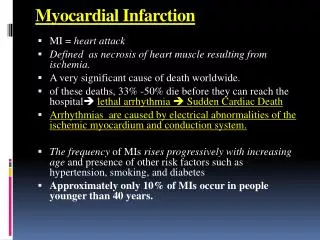
Myocardial Infarction. * Definition: coagulative necrosis of the myocardium due to acute coronary ischaemia . * Causes: T he m ost common cause is thrombosis on a preexisting disrupted atherosclerotic plaque e.g. hemorrhage, ulceration and rupture…. * Sites of MI:.


Myocardial Infarction
E N D
Presentation Transcript
* Definition:coagulative necrosis of the myocardium due to acute coronary ischaemia. * Causes: Themost common cause is thrombosis on a preexisting disrupted atherosclerotic plaque e.g. hemorrhage, ulceration and rupture…
* Sites of MI: I. Left ventricle (the commonest): • a. Anterior infarct (anterior wall of the Lt .ventricle + apex + anterior part of IV septum): due to occlusion of the anterior descending branch of the left coronary artery. • b. Lateral infarct (lateral wall of the Lt. ventricle): due to occlusion of circumflex branch of the left coronary. • c. Posterior infarct (posterior wall of the Lt. ventricle): due to occlusion of a branch of the Rt. coronary artery that supplies that part of the left ventricle. II. Right ventricle (rare): due to occlusion of the right coronary artery.
* Types of MI: • Transmural • Full thickness. • Subendocardial • Inner 1/3 of myocardium
MI: day 1, day 3, day 7
* Clinical Features of MI: 1. Pain: • Severe crushing substernal chest pain, which may radiate to the neck, jaw, epigastrum, shoulder or left arm. • Pain lasts for hours to days and is not relieved by nitroglycerin. • Absent in 20-30% of patients (diabetics, hypertensive, elderly). 2. Pulse is rapid and weak. 3. Dyspnea. 4. Cardiogenic shock in massive MI(>40%of lt. ventricle). 5. ECG shows typical findings of ischemia.
* Laboratory evaluation of MI: 1. Troponins: • The most specific test for MI. • Become detectable after 2 to 4 hours, reach the peak at 48 hours. Their levels remain elevated for 7 to 10 days. 2. Creatine kinase (CK-MB): • Is the second best marker, however, it increases in the blood also when there skeletal muscle damage. • It begins to rise within 2 to 4 hours of MI, peaks at 24 to 48 hours and returns to normal within approximately 72 hours. 3. Lactate dehydrogenase (LD1). • Rise 24 hrs, peaks 72 hrs, persists 72 hrs. • Non-specific because it aslo increases in cases of cancer, encephalitis, meningitis…
* Complications of MI: • Occurs in more than 90% of cases. • Cardiac arrhythmia: - Sudden death can occur due to ventricular arrhythmia. 2. Left ventricular failure. 3. Cardiogenic shock. 4. Myocardial rupture and hemopericardium. 5. Thromboembolism:Thecombination of a local myocardial abnormality in contractility (causing stasis) with endocardial damage (causing a thrombogenic surface) can foster mural thrombosis and, potentially, thromboembolism .
6. Pericarditis 7. Ventricular aneurysm. 8. Chronic heart failure.