Percivall Pott
Percivall Pott. 1714 – 1788 Chimney sweeps cancer of the scrotum Pott’s disease – vertebral tuberculosis. John Hunter. 1728 – 1793 Papers at the Royal Society on experimental pathology, including the use of a microscope Described inflammation

Percivall Pott
E N D
Presentation Transcript
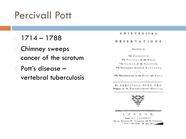
PercivallPott • 1714 – 1788 • Chimney sweeps cancer of the scrotum • Pott’s disease – vertebral tuberculosis
John Hunter • 1728 – 1793 • Papers at the Royal Society on experimental pathology, including the use of a microscope • Described inflammation • Hunterian Museum, Royal College of Surgeons, London
First Systematic Textbook of Pathology • Matthew Baillie – 1761 – 1823 • Nephew of John Hunter • The Morbid Anatomy of Some of the Most Important Parts of the Human Body • Microscopic pathology atlas • Physician of King George III
Thomas Hodgkin • 1798 – 1866 • On Some Morbid Appearances of the Absorbent Glands and the Spleen • “Lister’s compound microscope might lead to useful discoveries in the future.”
Joseph Recamier – 1774 – 1852 – metastasis • Richard Bright – 1789 – 1858 – Kidney disease • Thomas Addison – 1793 – 1860 – Pernicious anemia
Cell Theory • Robert Hooke – 1635 – 1703 – cell • Matthias Jacob Schleiden – 1804 – 1881 – botanist • Theodor Schwann – 1810 – 1882 – zoologist
Cell TheoryJohannes Peter Müller • 1801 – 1858 • Berlin • Father of medical microscopy • Microscopic criteria for benign and malignant tumors • Über den FeinernBau und die FormenderKrankhaftenGeschwülste– On the Finer Structure and Form of Morbid Tumors
Cell TheoryRudolph Virchow • 1821 – 1902 • The greatest figure in the history of Pathology • Die Cellularpathologie • “Omnis cellula e cellula” – all cells from cells
Herman Lebert – 1831 – 1878 • Microscopic atlas
1850s • Pathology developed as a separate specialty • Medical schools, Professors of Pathology • Microscope, diagnostic histopathology, neoplasia • France – laboratories • Germany – universities
Microscope • Fresh tissue, cut by hand, unstained • Formaldehyde fixation – Isaac Blum – 1833 – 1903 • Paraffin embedding – Edwin Klebs– 1834 – 1913 • Microtome – Minot – 1852 – 1914 • Biological stains • Hematoxalin – Franz Böhmer • Paul Ehrlich – 1854 – 1915
Anaplasia • David Von Hansemann • 1858 – 1920
Grading, Carcinoma in Situ • Albert Compton Broders • 1885 – 1964 • Mayo Clinic
Staging • Cuthbert Esquire Dukes • 1890 – 1977 • St. Mark’s Hospital, London
Pap Smear • George Papanicolaou – 1883 – 1962 • January, 1928 – New Cancer Diagnosis – Betterment Conference Battle Creek Michigan • 1941 – The Diagnostic Value of Vaginal Smears in Carcinoma of the Uterus • 1943 – Diagnosis of Uterine Cancer by the Vaginal Smear • 1954 – Atlas of exfoliative Cytology