Download
1 / 17
170 likes | 273 Vues
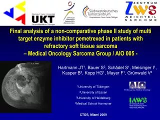
This study delves into the GABA_B receptor heteromeric complex, focusing on the unique functional attributes of the R1 and R2 subunits. Through analysis of intrinsic features such as signal peptides and transmembrane helices, we explore the underlying mechanisms of ligand binding and signaling. Additionally, we model human RYK and analyze phosphorylation sites to understand their effects on protein interactions. Utilizing homology modeling techniques, we demonstrate how known structures assist in the construction of accurate models, shedding light on critical biochemical processes.

E N D
Putting it all together to answer a “real” question Rob Russell Cell Networks University of Heidelberg
Domains assemble to form higher-order structures Pawson & Nash, Science, 2003
Case study 1: GabaB R1/R2 • Family 3 GPCRs • Subunit R1 binds ligands, R2 signals, but not vice versa • Why?
Analysis of intrinsic features Signal peptide Transmembrane helices Low complexity Coiled coil region PFAM analysis >gi|3776094|emb|CAA09940.1| GABAB receptor, subunit 1b [Homo sapiens] MGPGAPFARVGWPLPLLVVMAAGVAPVWASHSPHLPRPHSRVPPHPSSERRAVYIGALFPMSGGWPGGQACQPAVEMALEDVNSRRDILPDYELKLIHHDSKCDPGQATKYLYELLYNDPIKIILMPGCSSVSTLVAEARMWNLIVLSYGSSSPALSNRQRFPTFFRTHPSATLHNPTRVKLFEKWGWKKIATIQQTTEVFTSTLDDLEERVKEAGIEITFRQSFFSDPAVPVKNLKRQDARIIVGLFYETEARKVFCEVYKERLFGKKYVWFLIGWYADNWFKIYDPSINCTVDEMTEAVEGHITTEIVMLNPANTRSISNMTSQEFVEKLTKRLKRHPEETGGFQEAPLAYDAIWALALALNKTSGGGGRSGVRLEDFNYNNQTITDQIYRAMNSSSFEGVSGHVVFDASGSRMAWTLIEQLQGGSYKKIGYYDSTKDDLSWSKTDKWIGGSPPADQTLVIKTFRFLSQKLFISVSVLSSLGIVLAVVCLSFNIYNSHVRYIQNSQPNLNNLTAVGCSLALAAVFPLGLDGYHIGRNQFPFVCQARLWLLGLGFSLGYGSMFTKIWWVHTVFTKKEEKKEWRKTLEPWKLYATVGLLVGMDVLTLAIWQIVDPLHRTIETFAKEEPKEDIDVSILPQLEHCSSRKMNTWLGIFYGYKGLLLLLGIFLAYETKSVSTEKINDHRAVGMAIYNVAVLCLITAPVTMILSSQQDAAFAFASLAIVFSSYITLVVLFVPKMRRLITRGEWQSEAQDTMKTGSSTNNNEEEKSRLLEKENRELEKIIAEKEERVSELRHQLQSRQQLRSRRHPPTPPEPSGGLPRGPPEPPDRLSCDGSRVHLLYK Homology to known structure can be used to create model
Family III GPCRs Ligand binding domain R1 cut EC2 EC1 EC3 1 2 3 4 5 6 7 dimerisation IC2 IC1 IC3 Cterm - - - G-protein
R1 binds ligand R2 signals Robbins et al, J. Neurosci, 21, 8043, 2001
GabaB R1/R2 Ligand binding domain R2 R1 (none) EL2 EL1 EL3 EL2 EL1 EL3 1 2 3 4 5 6 7 1 2 3 4 5 6 7 - IL2 IL2 IL1 IL1 - - - IL3 Cterm IL3 Cterm blocked - - - G-protein
Human RYK model Human RYK (model) Insulin receptor YK (template) Katso, Russell, Ganesan, Mol Cell Biol, 19, 6427, 1999
Case study 3: What are phosphorylation sites doing? Van Noort et al, Mol Sys Biol, 2012
MPN134 is phosphorylated at Serine 392 Katso, Russell, Ganesan, Mol Cell Biol, 19, 6427, 1999
What do modifications do to interfaces? From polar to negatively charged From positively charged to polar Modelled MPN134 homodimer: From a polar-polar interaction to a pair of negative charges in proximity Van Noort et al, Mol Sys Biol, 2012
Homology modelling algorithm +
Homology modelling steps • Identify the homologue of known structure • Get the best alignment of your sequence to the structure • Model building • Side-chain replacement • Loop building • Optimisation/relaxation/minimisation
Problem with loops Two subtilisin-like serine proteases