Download
1 / 1
20 likes | 298 Vues
Dual mTOR/HDAC inhibition: preclinical development of a novel breast cancer therapy Mariya Yevtushenko, Kathleen A. Wilson- Edell , and Christopher C. Benz Buck Institute for Research on Aging, Novato, CA. Background. 2 . MLN0128 and TSA synergistically decrease breast cancer cell viability.

E N D
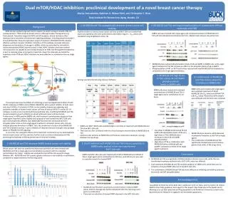
Dual mTOR/HDAC inhibition: preclinical development of a novel breast cancer therapy Mariya Yevtushenko, Kathleen A. Wilson-Edell, and Christopher C. Benz Buck Institute for Research on Aging, Novato, CA Background 2. MLN0128 and TSA synergistically decrease breast cancer cell viability 4. MLN0128 and TSA decrease phosphorylation of downstream effector molecules of mTOR HER2 (human epidermal growth factor receptor-2) and/or estrogen receptor (ER) are overexpressed in ~80% of human breast cancers. Although modern therapeutics (e.g. Trastuzumab, Tamoxifen) target the HER2 and ER receptors, clinical resistance often develops due to activation of downstream signaling pathways, including phosphoinositide 3 kinase-AKT/protein kinase B-mammalian target of rapamycin (PI3K/AKT/mTOR), despite effective upstream receptor inhibition. Activation of this pathway increases ribosome biogenesis and translation of oncogenic mRNAs, which are controlled by intracellular histone deacetylase (HDAC) activity as well. In fact, HDAC inhibitors that have entered clinical practice are capable of overcoming resistance to HER2 and ER targeted therapeutics, in part by inducing decay of oncogenic transcripts. Given this rationale, we tested the hypothesis that mTOR and HDAC inhibitors are more effective in combination than as single agents. Viability studies on various breast cancer cell lines and MCF-10A non-transformed mammary epithelial cells were performed as described in figure 1. IC50 values were calculated using GraphPad (La Jolla, CA). SKBR3 cells were treated with single agent and combined treatment of MLN0128 and TSA with the indicated concentrations for 8 hrs. Western blot analysis was performed. • MLN0128 alone reduced phosphorylation levels of S6 and 4eBP1 in SKBR3 cells, while single agent treatment of TSA did not have an effect on the phosphorylation of S6 or 4eBP1. • Phosphorylation of AKT on S473 was further reduced with dual MLN0128/TSA treatment as compared to the single agent treatments. 6. Combination of MLN0128 and TSA reduce polysome formation in SKBR3 cells 5. MLN0128 and high doses of TSA reduce S6 and 4eBP1 phosphorylation levels in SKBR3 cells Synergy was determined using Calcusyn Software SKBR3 cells were treated with single agent and combined treatment of 50nM MLN0128 (10x IC50 value) and 500nM TSA (16x IC50value). Polysome profile analysis was employed and fractions were collected. SKBR3 cells were treated with indicated concentrations of MLN0128 and TSA in single agent and in combination for 24 hours. This project examines the effects of combining a novel investigational inhibitor of both mTOR complexes (mTORC1 and mTORC2), MLN0128, with a potent inhibitor of both class I and class II HDACs, Trichostatin-A (TSA), on the viability, downstream signaling, and polysome assembly of human breast cancer cell lines of various (HER2-/+ and/or ER -/+) subtypes, as well as non-transformed breast epithelial cells. Combining MLN0128 and TSA caused synergistic growth inhibition in almost all breast cancer cell lines tested. Furthermore, in HER2-positive SKBR3 cells dual treatment caused greater apoptosis than single agent treatment, while viability and apoptosis of non-transformed MCF-10A cells were dramatically less affected. Dual MLN0128/TSA treatment decreased AKTS473 phosphorylation more so than single agent treatment in all breast cancer cells, and also reduced polysome formation in SKBR3. Thus, the mechanism of action of dual MLN0128 and TSA treatment involves, in part, inhibition of ribosome function through a two-pronged attack on PI3K/AKT/mTOR signaling. In summary, the synergistic effects of this treatment combination across phenotypically diverse breast cancer cell lines warrants further study and clinical development of this promising dual pathway inhibiting breast cancer treatment strategy. • SKBR3 and MCF7 NEO3 cells exhibited higher sensitivity to treatment with MLN0128 and TSA than MCF-10A cells. • There was no clear correlation with the clinical subtypes and sensitivity to MLN0128 and TSA. • Cell lines most sensitive to MLN0128 and TSA dose combination resulted in synergy between the two drugs. • Low doses of MLN0128 decreased S6 and 4eBP1 phosphorylation levels, while very high doses of TSA (1000nM) were necessary to block S6 and 4eBP1 phosphorylation. • High doses of TSA in combination with MLN0128 further inhibited pS6 and p4eBP1 compared to either of the single agent treatments. • MLN0128 alone caused a subtle decrease in polysome formation, as did TSA as single agents. • The combination of MLN0128 and TSA significantly blocked polysome formation, greater than either of the drugs alone. 1. MLN0128 and TSA decrease SKBR3 breast cancer cell viability 3. Dual treatment with MLN0128 and TSA induces apoptosis in SKBR3 cells, and not in the non-transformed MCF-10A cells Breast cancer cells and non-transformed mammary epithelial cells were treated with MLN0128 and TSA in single agent and combination treatment with the indicated concentrations for 3 days. Viability was analyzed by the CellTiter-Glo assay (Promega, Madison, WI). MLN0128 and TSA caused a greater reduction in cell viability in combination, compared to single treatment of either drug alone. Conclusions SKBR3 and MCF-10A cells were treated with 25nM of MLN0128 and 100nM of TSA in single agent and in combination for 48 hours, and Adriamycin was used as a positive control for cleaved PARP MLN0128 and TSA synergistically inhibited viability in breast cancer cells, while the non-transformed mammary epithelial cells, MCF-10A’s, were less affected. Dual treatment of MLN0128 and TSA induced apoptosis in SKBR3 cells, while there was no induction of cleaved PARP observed in the MCF-10A cells. The combination of MLN0128 and TSA was more effective at inhibiting cell viability, polysome formation, and AKT phosphorylation. Acknowledgements I would like to thank the entire Benz lab, in particular my PI, Dr. Benz, and my mentor, Dr. Wilson-Edell for their help, guidance, and support in this project. Also thank you to the Kapahi lab for allowing us to use their polysome profiling equipment, and Dominican University, as well as the Buck Institute for Research on Aging for this incredible opportunity. • MLN0128 and TSA alone caused only a small increase in cleaved PARP levels, and this cleavage was further enhanced when the two drugs were given in combination. • There was no induction of cleaved PARP observed in the MCF-10A cells.