Download
1 / 34
340 likes | 535 Vues
VKG-vid akuta koronara syndrom. Docent Sven V Eriksson E-mail: sven@sjukhus.org Hemsida : www.sjukhus.org. Docent Sven V Eriksson Åre 1999. History 1. ECG - (Waller 1887, Einthoven 1893) ST-elevation MI - (Smith 1918) Monocardiogram - (Mann 1920) AHA V1-6, 1938

E N D
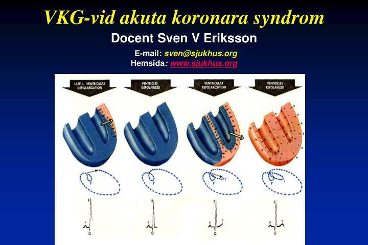
VKG-vid akuta koronara syndrom Docent Sven V ErikssonE-mail:sven@sjukhus.orgHemsida: www.sjukhus.org
Docent Sven V Eriksson Åre 1999
History 1 • ECG - (Waller 1887, Einthoven 1893) • ST-elevation MI - (Smith 1918) • Monocardiogram - (Mann 1920) • AHA V1-6, 1938 • 12-lead ECG, (Goldberger 1942) • VCG - (Schmitt 1955)
History 2 • Standard VCG-leads (Frank 1956/60) • VCG MI criteria (Hoffman 1964, Young 1968) • Continuous VCG (cVCG) (Hodges 1974) • cVCG during acute MI (Sederholm 1984) • On-line cVCG (Gröttum 1985) • MIDA 1986 • EASI-MIDA 2001
Myter 1 • VKG används nästan bara i Sverige • Det krävs ingen genuin kunskap i fysiologi för att lära sig VKG • Det är enkelt att inkludera VKG-analyser i t.ex. läkemedelsstudier • Av multiavlednings-EKG-metoderna är bara VKG utvärderade vid akut IHD • VKG används idag nästan bara i MIDA-systemen • Det krävs minst 8 avledningar för beräkning av VKG
Myter 2 • Icke-Q-vågs-infarkt = icke-transmural infarkt • Icke-Q-vågsinfarkt har annan prognos än Q-vågs infarkt • Icke-Q-vågs infarkt har icke ockluderat koronarkärl i motsats till Q-våginfarkt (22% skillnad). • Om cVKG är u.a. och pat. är smärtfri = ej MI • cVKG ger ingen information vid grenblock • Om cVKG/Holter visar ischemi = myokard-ischemi
Q Versus Non-Q • Prinzmetal. Q-wave=Transmural MI. Am J Med 1954;16:469-88. • Pipberger/Prinzmetal. Admitting errors of method ..”there was no reason to suppose that subendocardial infarcts could not generate Q-waves”. Am Heart J 1957;54:511-29. • Approximately 50 % of all subendocardial infarcts are accompanied by Q-waves. Circulation 1958;18:600-11, Circulation 1958;18:612-22. • Review of prognosis in non-Q versus Q in 9 studies. No difference! Table 1. JACC 1999;33:576-82.
Frank X-, Y- and Z-leads • X • Z • X • Y • Y • Z
S T x X S T y Y S T z Z • ST-VM • = • 2 • 2 • 2 • ST • + • ST • + • ST • x • y • z • ST-Vector Magnitude- ST-VM
S T S T - v e t o r c y Z S T z X S T x Y ST-Vector
P r e s e n t S T - v e c t o r Y S T C - V M Z I n t a S T - v e c t o r i i l X ST Change Vector Magnitude STC-VM
X Y A Z QRS-Vector Difference QRS-VD
Why use more/other than 12-lead ECG? • Matetzky S et al. Significance of ST Segment Elevations in Posterior Chest Leads (V7 to V9) in Patients with Acute Inferior Myocardial Infarction: JACC 1998;31:506-11. • Jai B et al. Importance of posterior chest leads in patients with suspected myocardial infarction, but nondiagnostic, routine 12-lead electrocardiogram. Am J Cardiol 1999;83:323-6. • Addition of right precordial leads to standard exercise electrocardiography improves sensitivity. N Engl J Med 1999;340:340-383.
Limits: • QRS-VD >15 uVs • ST-VM > 0.05 mV
QRS-VD känslig för: • Ändrat kroppsläge (ofta typisk bild) • Ischemi • Ändring i volym • Ledningshinder
ST-VM känslig för: • Ischemi (relativt spec./män) • Digitalis • Frekvens • Vänsterkammarhypertrofi
ECG/VCG difficult in patients with: • Bundle branch block? • Ventricular pacing • Left ventricular hypertrophy? • Atrial fibrillation?
VCG can give information regarding: • Ischemia (predischarge exercise test) • Prognosis (MI/Unstable angina) • Reperfusion • Reocclusion • Diagnosis (bundle branch block)
Value of clinical and VCG data for prediction of ST depression at exercise test X2 value P value STC-VM episodes 31.5 <0.001 ST-X maximum depression 16.2 <0.001 ST-Z value elevation 9.4 <0.01 Rest pain episodes 5.5 <0.05 Lundin P, Eriksson SV et al. J of Electrocardiol 1995;28:277-85
Prognostic information • Lundin P, Eriksson SV, Strandberg L, Rehnqvist N. Prognostic Information from on-line vectorcardiography in acute myocardial infarction. Am J Cardiol, 1994;74:1103-1108. • Lundin P, Eriksson SV, Fredriksson M, Rehnqvist N. Prognostic information from on-line vectorcardiography in patients with unstable angina pectoris. Cardiology, 1995;86:60-66. • Andersen K, Eriksson P, Dellborg M. Ischaemia detected by continuous on-line vectorcardiographic monitoring predicts unfavourable outcome in patients admitted with probable unstable coronary disease. Coron Artery Dis 1996;7:753-760. • Andersen K, Eriksson P, Dellborg M. Non-invasive risk stratification within 48 h of hospital admission in patients with unstable coronary disease. Eur Heart J 1997;18:780-788. • Holmvang L et al. Relative contributions of a single-admission 12-lead electrocardiogram and early 24-hour continuous electrocardiographic monitoring for early risk stratification in patients with unstable coronary artery disease. Am J Cardiol 1999;83:667-674.
Variable Age Performed ex-test VF during hospitalization STC-VM episodes QRS-end value VCG sign of reperfusion Dead (n=36) Alive (n=167) 73+7 63+10** 42% 92%** 19% 4%** 3(2-5) 0(0-2)** 25(17-36) 19(15-30)* 22% 46%* Comparison between 167 survivors and 36 non-survivors *p<0.05, **p<0.01 Lundin P, Eriksson SV et al. Am J Cardiol 1995;74:1103-08
Markers of reperfusion ECG Symptoms Serum markers On-line VCG/ECG Relief of pain a 5 point reduction on a 1 to 10 scale Abrupt increase of Troponin-T/I CK-MB Myoglobin Combination A “snapshot” > 50% reduction of of ST elevation % ST recovery Accuracy 80% 35-50% of patients have multiple periods of both ST recovery and reelevation, reflecting cyclic variations in infarct artery flow
Signs of reperfusion: • > 50% reduction of ST-VM within 90 min • Early “plateau” of QRS-VD
Actually open (n=12) 5 + collateral 1 - collaterals Actually Closed (n=19) 12 - collaterals 7 + collateral's VCG monitoring to assess early vessel patency Chest pain + Thrombolytic drug On-line VCG 90 min (n=96) “Open” by VCG (n=65) • Actually closed • (n=7) • Closed by VCG (n=31) Actually open (n=58) Dellborg et al. Eur Heart J 1995;16:21-29
Selected publications • Lundin P, Eriksson SV et al. Ischemia monitoring with on-line vectorcardiography compared with results from a predischarge exercise test in patients with acute ischemic heart disease. J of Electrocardiol 1995;28:277-285 • Lundin P, Eriksson SV et al. Ischemia monitoring with on-line vectorcardiography during dobutamine stress-echocardiography in patients with unstable coronary artery disease. J Int Med., 1998;244:61-70 • Lundin P, Eriksson SV et al. Ischemia monitoring with on-line vectorcardiography during dobutamine stress-echocardiography in patients with unstable coronary artery disease. J Int Med., 1998;244:61-70 • Jensen J, Eriksson SV et al. Systolic deterioration in basal segments of the left ventricle is related to myocardial ischemia during angioplasty: A tissue Doppler echocardiographic and vectorcardiographic study. Clinical Science 2001;100:137-143 • Jensen J, Eriksson SV, Lindvall B, Lundin P. Sylvén C. Women react with more myocardial ischemia and angina pectoris during elective percutaneous transluminal coronary angioplasty. Cor Art Disease 2000:11;527-35. • Eriksson SV. Vectorcardiography: a tool for the non-invasive detection of reperfusion and reocclusion? Thrombosis and Haemostasis 1999;82:64-67
VCG-studies DS/HS/USA/Germanywww.sjukhus.org for more details • VCG during acute MI 210 pat. DS, Thesis 1995 • VCG in unstable angina 160 pat. DS • PEGHIRUID 210 pat. DS “core-lab”, Berlin, Germany • VCG-registration during PTCA 209 pat. HS, Thesis 2000 • VCG during dialysis DS. 120 registrations • VCG-in Chest pain unit, 1918 pat. Chattanooga, USA • EASI/Frank-MIDA during PTCA 108 pat. • EASI/Frank-MIDA during thallium 90 pat.
The Erlanger/DS VCG-study Assistant Professor, Francis M. Fesmire UT College of Medicine Assistant Professor, Sven V Eriksson, Danderyds Hospital, Karolinska Institutet
Patients in VCG-study 2 206 consecutive pat 210 with BBB 2 128 with VCG 1 918 pat.
Characteristics in patients with and without LVH on ECG With LVH Without LVH N=196 N=1 722 Age 54.5 + 13.453.5 + 13.9 Male 99 (50.5%)889 (51.6%) Race Caucasian 86 (43.9%) 1326 (77.0%)*** African American 107 (54.6%) 379 (22.0%)*** Other 3 (1.5%) 17 (1.0%) Previous MI 65 (33.2%) 496 (28.8%) Previous PTCA/CABG 47 (24.0%) 410 (23.8%)
Conclusions: • VCG registration improves identification of patients with high risk of an acute MI • The optimal cut-off value for patients without left ventricular hypertrophy is 100 uV • In pat. with LVH, VCG-monitoring has limited power for detection of acute MI