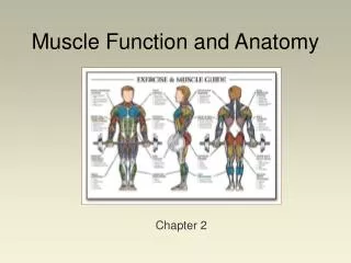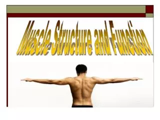
Muscle Structure and Function
Muscle Structure and Function. KIN 2711 March 6, 2006. Muscle Types. Three types of Muscle Skeletal Cardiac (myocardium) Smooth key differences - appearance, structure innervation, fuel supply. Muscle Function. Produce movement Maintain postures and positions Stabilize joints

Muscle Structure and Function
E N D
Presentation Transcript
Muscle Structure and Function KIN 2711 March 6, 2006
Muscle Types • Three types of Muscle • Skeletal • Cardiac (myocardium) • Smooth • key differences - appearance, structure innervation, fuel supply
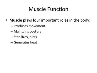
Muscle Function • Produce movement • Maintain postures and positions • Stabilize joints • Usually for other muscle activity • Support and protect viscera • Control cavity pressures • Produce heat to maintain body temperature • Control entrance and exits to body • swallowing, defecation, urination • Energy Storage
Organizational Hierarchy • Skeletal muscles have a hierarchical structure • the muscle (organ) is composed of fascicles which are composed of fibres • a fibre is a single cell so the term is used interchangeably • Note that muscle cells are multinucleated
Organizational Hierarchy • Fibres are composed of myofibrils which are arranged in parallel (side by side) • myofibrils are composed of sarcomeres in series (end to end) • sarcomeres are the functional unit of the muscle cell • are composed of think and thin myofilaments
Sarcomere • If one looks at muscle with a light microscope then one finds cross-striations of the sarcomeres • tell about the structure of the sarcomere
Physiological Classification • Burke and coworkers classified the motor units in cat muscle based on three factors • 1. Contraction time • 2. “sag” in force during unfused tetanus • 3. Fatigue test • Resulted in Slow, Fast Fatigue-Resistant, and Fast Fatigable
Motor Units • Consists of a cell body, an -motoneuron, and all the muscle fibres innvervated by that -motoneuron • so when the -motoneuron is stimulated then all the fibres in that motor unit will contract • number of fibres in a given motor unit will vary according to degree of motor control • those with a fine control (like in the hand) will have few fibres (10 to 100) while those in the leg will have 1000s
Neuron Anatomy • -motoneuron goes to the muscle cell
Size Principle of Motor Unit Recruitment • Frequency of motor unit use is related to the size and the ease of triggering an action potential in the soma • Key Points • the smaller the soma (neuron cell body) the easier to recruit • slow units have smaller bodies • therefore slower units will be recruited first • then the larger faster units
Recruitment Patterns • This works well • Why? • Smaller more efficient units come into play first • more accurate matching between workload and power output by the cell
EMG • Electromyography can be used to measure the recruitment of motor units in the muscle • EMG measures both frequency and amplitude of electrical activity • gives idea of type and number of motor units • if coupled with force measurements then a relationship between force and EMG can occur
Rate Coding • Information is sent through a nerve by rate coding • higher frequency signal, higher force production • works for single muscle, single motor unit, a single fibre
Muscle activity in cycling • EMG data has been collected by numerous researchers • measured surface activity then normalized signal to max isometric voluntary contraction
EMG activity • Electromyography - measures electrical signal produced by contraction • bi-polar electrodes are placed on the skin around the motor point (where major nerves enter into muscle in question) • measures depolarization followed by repolarization
EMG • The signal does not correlate exactly with force development • period of electromechanical delay • note that tension has to produced first to stretch the elastic components in muscle before the force is transmitted to the bones
EMG • Can be analyzed in a number of ways • raw data • rectified - all signals shown as positives • integrate over time period and reset - to be able to quantify total emg signal
EMG • EMG has been used to demonstrate different way s of force development (A, B) • also used to demonstrate a muscle’s response to activity
Rectus Femoris Hamstrings
EMG in cycling • Much room for more research • smaller muscles such as adductors, calf muscles have not been examined • secondly, surface EMG may not reflect what is going on in deep muscles • researchers have assumed that crank velocity is constant
Other Means to Increase Force Production • Can also recruit more motor units • forces from each motor unit are additive
Contractile Properties • Contraction refers to the activation of the force generating activities of the muscle • during a contraction, the muscle can shorten, lengthen or not move at all • No Change in length = isometric • Shortening = Concentric or Miometric • Lengthening = Eccentric, Pliometric
Length-Tension Relationship • Muscle fibre has an optimal length • amount of force it can develop varies with that length
Storage and release of Elastic Energy in the Locomotor System Stretch-Shortening Cycle
Elastic Energy • Elastic Energy is very important in terms of movement • Energy that is stored and released • Improves jumping performance • Reduces amount of energy expenditure by muscles ( less use of fuel) during walking and running • Reduces impact during walking and running, i.e. fat pads in feet
Principles of Elasticity • Think of elastic material as a cylinder • If stretched, area Ao decreases to A and length lo increases to l • Slope k in graph c is the stiffness of the material (steeper the slope the more stiff) • The amount of work is the shaded area
Principles of Elasticity • Properties • Depend on dimensions and nature of materials • Think of steel and rubber
Tensile Stress • How much stress can a tendon take? 1) true tensile stress • F/A – not convenient because are has to test material to see how much more slender it gets as it stretches 2) nominal tensile stress • F/Ao • Note - very little differences if A-Ao is small • In case of Tendon – small extension don’t effect x-sectional area -- up to 8-10% before break
Properties of Body Parts • Tensile tests on gastrocnemius of walking • Stretched and relaxed at certain rate – like hopping strides • Peak force of 700N • Same peak force as hop by wallaby • Only ran machine a few times – to avoid ‘creep’
Gastrocnemius • Area under the graph is work • Note that going up is stretching – energy in • And going down is shortening – energy out Loop is energy that is lost – usually as heat • Energy Dissipation • For most mammalian tendons most of the energy is returned • About 88 to 95%
Energy Dissipation • Should be low • Energy lost could have been used for movement • Lost heat – in horses galloping tendon temperature can reach 45C in leg tendons • Bad – energy needs to be used for cooling rather than movement – sweating – blood flow shift • Proteins begin to change shape above 40C – become denatured
Ligaments and Fat Pads • Under heel in foot, there is a large thick fat pad • In the foot, there are a number of ligaments that act as springs • Both structures absorb shock and also act as energy return devices
Running v. Standing • Barefoot running films show a flattening of the arch • When pictures of feet resting lightly on the ground are compared to running the ankle is 10 mm closer to the ground during running
Ligaments • Forces in the foot • At running stage where forces are highest, a 70kg person have 2.7x BW on the foot • 1.9kN upwards force, and a 4.7kN force in the achilles means that the joint is undergoing a force of 6.4kN (1.9 + 4.7)
Ligaments • Foot is flattened • Ligaments are stretched • Plantar aponeurosis, long and short plantar ligaments and spring ligaments • Remember loop data in tendon • For entire foot the loop is wider, suggesting more energy loss, about 22% (energy returned would be 78%)
Heel Pad • Consists of collagen and fat • Energy dissipation calculated in 2 ways • Dissected out and squeezed in dynamic machine – 30% of energy lost • In a live subject, had pendulum swing down and hit heel – measure rebound – 75-95% • Why difference?
Heel Pad • Why? • if heel pad dissected out and hit with pendulum – 66% • Foot and leg not a rigid structure, so energy is lost elsewhere from side to side shifting of ankle • Concept of ‘creep’ – single contact gives different results than repeated contacts • 10 contacts results in dissipation going 48 to 32% Conclusion –30% energy dissipation --9 mm compression
Springs Functioning as Catapults • In a catapult or bow, stretch spring back • When bow is fired, string recoils rapidly firing arrow much faster than you could throw it • Energy returned in recoil equals work done to stretch bowstring but is returned at a greatly increased rate • Tendons do same in body
Force-velocity in muscle • Individual muscle has fibres that contract at many different speeds • In soleus of horse can vary between 0.3 to 3 fibre lengths per second • Max. shortening is determined by fastest fibres • Elastic recoil of tendon is not limited • Therefore no limit on speed of movement