Download
1 / 152
1.68k likes | 2.73k Vues
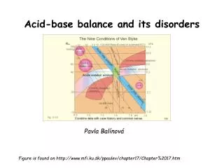
Chapter 4 Acid-base balance and acid-base disorders. Department of Pathophysiology, the School of Medicine, Shandong University 薛冰. internal environment homeostasis. Water balance. homeostasis. electrolyte balance. acid-base balance. Contents. 1. Acid-Base Balance

E N D
Chapter 4 Acid-base balance and acid-base disorders Department of Pathophysiology, the School of Medicine, Shandong University 薛冰
internal environment homeostasis Water balance homeostasis electrolyte balance acid-base balance
Contents • 1. Acid-Base Balance • Acid-base Biochemistry • Regulation of pH • Laboratory Tests • 2. Simple Acid-base Disorders • Metabolic Acidosis • Respiratory Acidosis • Metabolic Alkalosis • Respiratory Alkalosis • 3. Mixed Acid-base Disturbance
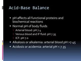
Part I:Acid-base balance and its regulation • The basic meaning of acid-base balance is the stable [H+] in the body fluid.
I、Concept of acid and base • acid: An acid is a H+ donor, when it is dissolved in water. After the loss of H+, it becomes a base. • HA (acid) → H+ + Aˉ(base) • H2CO3 → H+ +HCO3ˉ • H2SO4, H3PO4 • base: A base is a H+ acceptor, when it is dissolved in water. After the combining of H+,it becomes an acid • Aˉ(base)+ H+ → HA (acid) • HCO3ˉ+H+ → H2CO3 • OH-, HCO3-, SO42-, HPO42-, NH3
(I) Source of acid II、Sources of acid and base The main origin of acid and base is the intracellular metabolism (catabolism of protein, carbohydrate and fat). • volatile acid • fixed acid
1.volatile acid—— H2CO3 metabolism of protein, carbohydrate and fat H+ + HCO3- CO2 + H2O H2CO3 CA daily production:300-400L/d Excretion:lung Reabsorption in kidney RBC、kidney tubulesepithelium 、alveolar epithelial cell 、gastric mucosa
2. unvolatile acid (fixed acid):(50-100mmol/d) • Uric acid, phosphoric acid (H3PO4) and sulfuric acid (H2SO4) are the products in the metabolic process of proteins and nuclear acids. • Lactic acid and ketonic bodies (β-hydroxybutyric acid and acetoacetic acid) can be formed from the metabolic process of carbohydrate and fat as intermediate products, when the oxygen supply is not sufficiency. • Exogenous acid(food and drug): • Excretion through kidney
(II) Sources of base • Origin of bases • Endogenous: deamination―>NH3 Less than acid production • Exogenous input: vegetables, and fruits
[HCO3-] pH=pKa+lg [H2CO3] 20 =pKa+lg 1 Henderson-Hasselbalch Equation • Acid-base balance is mainly the balance between production and loss of acid and base. = 6.1 +1.3 = 7.4
Acid-base balance: pH∝ [HCO3-] / [H2CO3]orpH∝ [HCO3-] / PaCO2 [H+]↑ Buffer Respiratory Renal Excrete H+ Keep NaHCO3 paCO2↓ ECF ICF & bone Neutralize H+ eH+ & iK+exchange (Immediately) ( 2~4h) (1~3min) (hs;1~3d)
Buffer system Source Renal Respiratory Cellular
表1 全血五种缓冲系统 表2 全血中各缓冲体系的含量与分布 缓冲酸 缓冲碱 缓冲体系 占全血缓冲体系% H2CO3 HCO3¯+ H+血浆HCO3¯ 35 H2PO4- HPO42 ¯+ H+红细胞内Hb 18 HPr Pr ¯+ H+ HbO2-及Hb- 35 HHb Hb¯ + H+磷酸盐5 HHbO2 HbO2¯ + H+血浆蛋白7 (I)blood buffer system Buffer systems:consists of a weak acid and its’ salt ※HCO3-/H2CO3 is the most important buffer pair.
HCO3-/H2CO3 buffer system • Regulate CO2 or HCO3- through kidney and lung, the most important buffer pair(50%)。 • fixed acid and base buffer system • PH is dermatied by HCO3-/H2CO3。
hemoglobin buffer system (Hb-/HHb、HbO2-/HHbO2) character: RBC specificity volatile acid buffer CO2 CA:carbonic anhydrase CO2+H2O CA H2CO3 CA H+ HCO3- Cl- (RBC) HCO3- Cl- ← HHO2 HbO2- HHb Hb-
phosphate buffer system • HPO42-/H2PO4- character: play a role in cell and kidney
protein buffer system • Pr-/HPr Intracellular buffer
Mechanism of buffer HCl+NaHCO3→NaCl+H2CO3→CO2+H2O NaOH + H2CO3→NaHCO3 + H2O Accept H+or release H+,decrease the change of pH
Character of Buffer • Unvolatile acid: • HCO-3/H2CO3 system: • ½ of the buffer capacity • Opened regulation: respiratory and renal • Volatile acid : • Hb-/HHb、HbO2-/HHbO2
(II)Mechanisms of respiratory control change the depth or rate of respiration→change CO2 elimination→ [HCO3-]/ PaCO2→Acid-base balance
1. central chemoreceptor • PaCO2 (N:40mmHg) ↑→ pH of CSF↓ →tostimulatecentral chemoreceptor →☆ the respiratory center→Pulmonary ventilation volume↑ PaCO2 >60mmHg (8kPa) → Pulmonary ventilation volume↑10 times but, PaCO2 >80mmHg (10.7kPa)→inhibit respiratory center,named as carbon dioxide narcosis
central chemoreceptor The central chemoreceptor is sensitive to the change of CO2, which is easy to cross the blood-brain barrier. It takes time for the H+ to penetrate across the blood brain barrier into the interstitial fluid of the brain, the increase of [H+] in the brain is relatively slow, so the effect of H+ on the central chemoreceptor will be slow.
2.peripheral chemoreceptor • PaO2↓ 、pH ↓ 、PaCO2↑→tostimulateperipheral chemoreceptor →☆ the respiratory center→ Pulmonary ventilation volume↑ • PaO2﹤60mmHg (8kPa) → ☆ the respiratory center; but PaO2<30mmHg →inhibit respirator center。 • Less sensitive than central chemoreceptor
3.Characteristic of respiratory compensation (a) Timeliness. The respiratory response begins within several minutes. The respiratory response often takes 30 minutesfor the respiratory compensation. 12~24 hours to get maximal compensation. (b) limited compensation
(III) Renal regulation of acid-base Balance Renal compensation begins from several hours after the addition of acid load, and it may take 3~5 days to reach the maximum of this compensatory capacity. Kidneys play a major role in the regulation of pH in the body.
Excrete the nonvolatile acid, reabsorb the bicarbonate,“排酸保碱” →keep [HCO3-]→maintain acid-base balance。 HCO3- filtrate through glomerulus freely(5000 mmol/d),85%~90% is reabsorbed by proximal tubule,others are reabsorbed by distal convoluted tubule and collecting duct,0.1% is excreted→urine pH 6.0。 urine pH vary from 4.4 to 8.0
1.in proximal tubule (a) Na+-H+ exchange
2.in distal tubule & collecting duct α-intercalated cell: secrete H+ upper membrane: (a) H+-ATPase; (b) H+-K+ ATPase Urinary acidification (H2PO4-↑NH4+↑) base membrane: Cl- /HCO3-exchange
4. competitive inhibition between K+-Na+ exchange andH+-Na+ in distal tubule • K+-Na+ exchange: secrete K+, reabsorb Na+, • H+-Na+ exchange:secrete H+,reabsorb Na+ • acidosis, H+-Na+ exchange↑→ K+-Na+ exchange↓→hyperkalemia。
(IV) Cellular regulation • (a) H+-K+ exchange • (b) Cl-- HCO3- exchange • (c) Utilizing of bone salt • (d) Synthesis of urea from NH3
1. H+-K+ exchange When [H+] in ECF (serum) is increased, the H+ will move into the cells, as a exchange for electrical neutrality, K+ will shift from ICF to the ECF. So the pH of ECF (serum) will increase to normal, but hyperkalemia may occur.
2. Cl-- HCO3- exchange • When CO2 in ECF (serum) is increased, CO2 will move into the cells, CO2 combines H2O to form carbonic acid, then H2 CO3 dissociates to form H+ and HCO3¯ , the HCO3¯ moves out of the RBC, for neutrality, Cl ¯ moves into the cells.
3.Utilizing of bone salt • In chronic metabolic acidosis, bone salt, Ca3(PO4)2, is also utilized as a buffer base, but the expense is decalcification of bone and osteoporosis (loose and soft bone). • Ca3(PO4)2 + 4H+ ←→ 3 Ca2+ + 2 H2PO4 ¯ • It is not a good way of regulating acid-base balance by utilization of bone salt.
Buffer system Source Renal Respiratory Cellular
Part II laboratory tests of acid-base disturbances
1. pH • pH is the negative logarithm (-log) of [H+] in a solution. [H+]=40nmol/L (pH=7.4) • The normal range in artery blood =7.35~7.45 (7.41) • The survival range of pH=6.8~7.8 • According to the Henderson-Hasselbalch equation: The pKa is the dissociation constant of carbonic acid (=6.1)
24 [HCO3¯ ] metabolic factor pH =6.1+ log --------------------------------------- 1.2 [H2CO3] respiratory factors 20 = 6.1+ log---------- =6.1+1.3=7.4 1 The pH is determined by the ratio of [HCO3¯ ] 20 --------------=--------- [H2CO3] 1 No matter how the absolute amounts of HCO3¯ and H2CO3 change, once the ratio remains 20/1, the pH would be 7.4 (normal).
24 [HCO3 ¯ ] metabolic factorpH =6.1+ log -------------------------------------------- 1.2 [H2CO3]respiratory factors • The primary changes determines the nature of the acid-base imbalance. • The purpose of secondary change is to restore the pH. • According to the pH: • compensatory acid-base disturbances • decompensatory acid-base disturbances
Clinical significance of PH (anticoagulant artery blood, insulation of air) • A normal range of pH may represent three different situations: • ① acid-base balance; • ② compensatory acidosis or alkalosis; • ③ a mixed decompensatory acidosis and decompensatory alkalosis.
Clinical significance • pH<7.35 decompensatory acidosis • ( acidemia ) • pH>7.45 decompensatory alkalosis • (alkalemia)
2.PaCO2 (partial pressure of carbon dioxide in arterial blood) CO2 in blood: (a) 23% HbCO2 in RBC (b) 70% HCO3- in plasma (c) 7% CO2 molecule in plasma CO2 is determined by the rate of CO2 production and the rate of CO2 elimination. PaCO2 is the tension of CO2 caused by CO2 molecule movement. The normal range = 33~46(40) mmHg (4.39~6.25 kPa).
PaCO2 The capability of normal lung to eliminate CO2 is very good. CO2 retention will not occur with normal ventilation. Generally speaking, the PaCO2 is determined mainly by the respiration, so the PaCO2 is called the “respiratory factor”. Higher PaCO2 is due to the inhibition of respiration. Lower PaCO2 is due to overventilation.
Significance PaCO2>46mmHg Primary increase: respiratory acidosis Secodary increase: metabolic alkalosis (compensated by lung) PaCO2<33mmHg Primary decrease: respiratory alkalosis Secodary decrease: metabolic acidosis (compensated by lung)
3.[HCO3-] Actual bicarbonate (AB) The normal [HCO3¯ ] is 22~27(24) mmol/L. AB is measured under “actual condition” in which both respiratory factor and metabolic factor affected the [HCO3¯ ]. CO2 +H2O=H2CO3=H++HCO3 ¯
Standard bicarbonate (SB) • SB is measured under “standard condition” (temperature 37~38℃, full oxygenation of hemoglobin, PaCO2 = 40 mmHg). Standard condition means that the respiratory factor is eliminated, then the [HCO3¯ ] is only affected by metabolic factor. • Higher SB means metabolic alkalosis or respiratory acidosis compensated by kidneys. • Low SB means metabolic acidosis or respiratory alkalosis compensated by kidneys.
Normally the AB=SB. CO2 +H2O=H2CO3=H++HCO3- If AB>SB (CO2 retention), the reason must be the effect of respiratory factor, which indicates respiratory acidosis or metabolic alkalosis compensated by lung. If AB<SB (CO2 depletion), the reason must be the respiratory factor, which means respiratory alkalosis or the metabolic acidosis compensated by lung.
4.Buffer base (BB) • Sum of all buffer basees in blood • In plasma: HCO3 ¯ =24 • Protein¯ =17 • In RBC: Hb¯ • HbO2¯ =6.3 • HPO4 2¯ =1.0 • BB=45~55 mmol/L • Determined by metabolic factors