A Flexible, Open, Decentralized System for Digital Pathology Networks
260 likes | 387 Vues
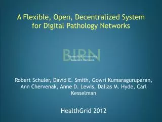
This paper explores a novel framework for digital pathology networks aimed at enhancing collaboration and data sharing among pathologists. With the rapid advancements in technology, digital pathology is transforming disease study and diagnosis, utilizing high-resolution whole slide images. The proposed system addresses challenges such as image size, format standardization, and security while providing features for real-time access, rich annotation, and distributed data management. By creating an open structure that emphasizes flexible data schemas and ontologies, the network promotes enhanced workflows and educational opportunities.

A Flexible, Open, Decentralized System for Digital Pathology Networks
E N D
Presentation Transcript
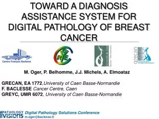
A Flexible, Open, Decentralized System for Digital Pathology Networks Robert Schuler, David E. Smith, Gowri Kumaraguruparan, Ann Chervenak, Anne D. Lewis, Dallas M. Hyde, Carl Kesselman HealthGrid 2012
Introduction • What is Pathology? • Study and diagnosis of diseases • Microscopy is a critical component • What is Digital Pathology? • Management of pathology data using computers • Emerging technology transforming sciences • What is a Whole Slide Image? • High-resolution capture of microscope slide (20x to 100x) • What is a Digital Pathology Network? • Sharing pathology data outside local environment • Platform to improve workflows, education, and collaboration
Motivation • Collaboration • Share pathology data across sites • Allow users to search across the entire participating network from a single request • Imaging • Instant access to whole slide images over the web • Rich annotation capabilities • Minimal client-side software • Security • Local storage and data entry • Control over which data to publish
Challenges - Collaboration • Application data schema and ontology differences • e.g. immunology research of animals vs. clinical oncology of humans • Differences even between labs within single organization • Requirements change as pathologists gain experience with the system • Proprietary and closed architectures • Limited data sharing • Difficult to integrate applications together • Distributed system with flexible data schema and ontologies to easily share data
Challenges – Image Size • Digital slide images are MUCH larger than typical digital images • Low network bandwidth • Images must be instantly viewable without full download 80,000 megapixels 40 GB each 14 megapixels 4 MB each 1/4th megapixel 300 KB each
Challenges – Image Format • No standard data or whole slide image format • DICOM supplement 145 for Whole Slide Imaging published but not yet implemented • Multiple closed image formats • Pixel storage and compression differences • Annotation storage, capabilities, coordinates • Metadata locations and implementation • Vendor-specific imaging clients • Limited platform, browser support • Feature-rich • Pixel data, annotations, and metadata must be converted to a common format
Challenges - Security • Sensitive data sets • Regulatory requirements • e.g. HIPAA • Protect access to data • Even to other parts of the organization • Network firewalls • Retain control over local environment • Data must be protected, maintained locally, and selectively published
Related Systems • Online digital pathology resources • Pathbase mutant mouse database • Zebrafish Atlas • Standalone virtual slide servers • OMERO No reusable infrastructure for digital pathology networks • Specimen Inventory Management • caTissue • Neuroimaging Systems • Human Imaging Database (HID) • eXtensibleNeuroimaging Archive Toolkit (XNAT) No whole slide image and annotation sharing • Scanner Vendor Systems • Olympus OlyVIA • AperioImageScope Limited platform and image support
Architecture Overview Pathology Information System (A) Site A Site B Data Access Service (B) Data Access Service (A) Data Integration Service Site A Pathology Information System (B) Site B
Architecture Overview Scan images at local centers Import Pathology Information System (A) Site A Site B Data Access Service (B) Data Access Service (A) Data Integration Service Site A Pathology Information System (B) Site B Import
Architecture Overview Curate data at local centers Pathology Information System (A) Data Entry Site A Site B Data Access Service (B) Data Access Service (A) Data Integration Service Site A Pathology Information System (B) Site B Data Entry
Architecture Overview Push published data to access services Pathology Information System (A) Site A Site B Transfer Data Access Service (B) Data Access Service (A) Data Integration Service Transfer Site A Pathology Information System (B) Site B
Architecture Overview Search for data across all sites Pathology Information System (A) Site A Search Site B Data Access Service (B) Data Access Service (A) Data Integration Service Query B Query A Site A Pathology Information System (B) Site B
Pathology Information System Secure Network at Local site Image Metadata Image Processing Service Pathology Workbench Import Whole slide images Scan Publish Curate Demilitarized Zone (DMZ) at Local Site
Pathology Information System – Image Processing Service Thumbnail Image Viewer Tiles VSI Original Image NDPI Common Annotations Image Processing Service SVS Acquisition Metadata
Pathology Information System – Pathology Workbench Graphical Annotation Spreadsheet Local Search Data entry workflow High-resolution Imaging Web Service Data Publication Pathology Workbench Image Metadata Whole slide images User Store
Pathology Information System – Pathology Workbench • Secure • Local access and data administration • One per center • Authenticate with username and password • Supports common credential store (LDAP) • Application and image-level permissions • Flexible, complex data schema and ontologies • Based on SNOMED • Data entry methods • Workflow-based web pages • Spreadsheet import • Web-service (REST)
Pathology Information System – Pathology Workbench • Zoomify Enterprise Viewer • Image viewing • Browser-based, loads instantly • Progressive-rendering • View whole image at low resolution • Zoom to high resolution area • Annotations • Mark, label points of interest • Share with other users
Pathology Information System – Pathology Workbench • Search local data • Specific metadata • e.g. find all images that belong to a lung specimen • Full-text • e.g. find all images that contain the phrase “Ringworm” in all metadata • Publication • Choose the data that gets pushed to collaborators
Data Access Service Data Access Service Image Metadata Whole slide images • Operated outside the firewall (DMZ) • Receives data securely from a single Pathology Information System • e.g. using Globus File Transfers (GridFTP) with host or service certificates • Manages its own copy of metadata and image files
Data Integration Service Example: Search the network for all images related to “Heart Disease” Data Access Service 1 User Query Data Access Service 2 Sub-query 1 Query Mediator Data Access Service N Sub-query 2 Sub-query N
Data Integration Service All “Heart Disease” images from all sites Data Access Service 1 Data Access Service 2 Result 1 Query Mediator Data Access Service N Result 2 Result N
Testbed – Initial Deployment • Two National Primate Research Center (NPRC) sites • 27,000+ images processed • Olympus VSI, Hamamatsu NDPI, Aperio SVS, JPEG, TIFF • Rate of conversion ~1 GB/hour per compressed image data • Acceptable for processing archives • Not ideal for instant access to 20+ GB slide from scanner, but okay for working with pre-defined sets • Processing images simultaneously for better capacity • Collection is over 1.5 TB in size • Pathologists curating data and assigning to images • Currently publishing data to share with other NPRC sites
Testbed – Data Schema • Developed over 18 months • Identified concepts and relationships • Rapid database implementation based on schema • Continual feedback and improvements • Combination of standard and custom ontologies • e.g. SNOMED
Future Work • Expand testbed beyond initial deployment • More National Primate Research Centers to collaborate in their network • New collaborative networks • Human pathology domain • Clinical use cases • Improve data curation experience • Further data schema refinement • Federate between different schemas • Image processing automation improvements • Scalability • Performance • Management
Conclusion • Digital Pathology Network system architecture and implementation met user requirements • Flexible, open, decentralized • High-resolution whole slide imaging with annotations • Complex pathology schema and data workflows • Local administrative control • Share data with collaborators • Successful initial deployment • Two large-scale research centers • Active use by pathologists and related research and support staff • Learn more about BIRN Pathology http://www.birncommunity.org