Download
1 / 131
1.35k likes | 1.59k Vues
Diseases of Blood Cells. Back to Basics. Blood is a liquid tissue A mixture of cells and water The water contains Protein, glucose, cholesterol, calcium, hormones, metabolic waste and hundreds of other substances

E N D
Back to Basics • Blood is a liquid tissue • A mixture of cells and water • The water contains • Protein, glucose, cholesterol, calcium, hormones, metabolic waste and hundreds of other substances • Plasma is the liquid portion of the blood containing the blood clotting protein Fibrinogen • Serum is the fluid remaining after the blood clots • Does not contain Fibrinogen
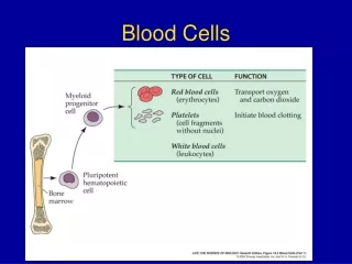
plasma (55%) red blood cells (5-6-million /ml) white blood cells (5000/ml) platelets
Plasma liquid part of blood plasma transports:- • soluble food molecules • waste products • hormones • antibodies
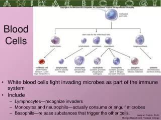
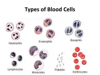
Production of Erythrocytes • Hematopoiesis – blood cell formation • Occurs in the red bone marrow (myeloid tissue) • Axial skeleton and girdles • Epiphyses of the humerus and femur • Marrow contains immature erythrocytes • Composed of reticular connective tissue • Hemocytoblasts give rise to ALL formed elements • Lymphoid stem cells - give rise to lymphocytes • Myeloid stem cells - give rise to all other blood cells
Production of Erythrocytes: Erythropoiesis • A hemocytoblast is transformed into a committed cell called the proerythroblast • Proerythroblasts develop into early erythroblasts • The developmental pathway consists of three phases • Phase 1 – ribosome synthesis in early erythroblasts • Phase 2 – hemoglobin accumulation in late erythroblasts and normoblasts • Phase 3 – ejection of the nucleus from normoblasts and formation of reticulocytes • Reticulocytes then become mature erythrocytes • Reticulocytes make up about 1 -2 % of all circulating erythrocytes
Regulation and Requirements for Erythropoiesis • Circulating erythrocytes – the number remains constant and reflects a balance between RBC production and destruction • Too few red blood cells leads to tissue hypoxia • Too many red blood cells causes an undesirable increase in blood viscosity • Erythropoiesis is hormonally controlled and depends on adequate supplies of iron, amino acids, and B vitamins
Hormonal Control of Erythropoiesis • Erythropoietin (EPO) released by the kidneys is triggered by: • Hypoxia due to decreased RBCs • Decreased oxygen availability • Increased tissue demand for oxygen • Enhanced erythropoiesis increases the: • RBC count in circulating blood • Oxygen carrying ability of the blood
Erythropoietin Mechanism Imbalance Start Normal blood oxygen levels Stimulus: Hypoxia due to decreased RBC count, decreased availability of O2 to blood, or increased tissue demands for O2 Imbalance Increases O2-carrying ability of blood Reduces O2 levels in blood Erythropoietin stimulates red bone marrow Kidney (and liver to a smaller extent) releases erythropoietin Enhanced erythropoiesis increases RBC count
Dietary Requirements of Erythropoiesis • Erythropoiesis requires: • Proteins, lipids, and carbohydrates • Iron, vitamin B12, and folic acid • The body stores iron in Hb (65%), the liver, spleen, and bone marrow • Intracellular iron is stored in protein-iron complexes such as ferritin and hemosiderin • Circulating iron is loosely bound to the transport protein transferrin
Platelets if you get cut:- • platelets produce tiny fibrin threads • these form a web-like mesh that traps blood cells. • these harden forming a clot, or "scab." • 150,000 to 400,000 per mm3
Thrombopoiesis • Thrombocytes or platelets • Derived from the megakaryocyte • Thrombopoietin • Formed in the liver and is similar to erythorpoeitin • Megakaryocytes fragment to form platelets • The spleen takes up about 1/3 of the platelets • Forms red plulp
Hemostasis = Opposite of hemorrhage stops bleeding Too little hemostasis too much bleeding Too much hemostasis thrombi / emboli Three major steps: • Vasoconstriction • Platelet plug Temporarily blocks the hole • Platelet-derived cytokines further the process • Coagulation cascade (= clot formation seals hole until tissues repaired) • Two pathways: Extrinsic and Intrinsic • After vessel repair, plasmin dissolves the clot
Steps of Hemostasis Vessel damage exposes collagen fibers Platelets adhere to collagen & release factors local vasoconstriction & platelet aggregation + feedback loop decreased blood flow platelet plug formation
Steps of Hemostasis cont. • Two coagulation pathways converge onto common pathway • Intrinsic Pathway. Collagen exposure. All factors needed are present in blood. Slower. • Extrinsic Pathway. Uses Tissue Factors released by injured cells and a shortcut. • Usually both pathways are triggered by same tissue damaging events. • The different factors can be subject to a variety of problems • Hemophilia • Hypercoagulable states
The Role of the Platelet • VIII – Von Willebrand Factor • vWF can bind to exposed collagen and also to receptors on the surface of platelets • Platelets are delivered to the damage site and bind to vWF = platelet activation • Platelet Plug • Platelets expose receptors • Degranulation – release of ADP to promote platelet activation (pa) • Releases Thromboxane A2to promote pa
Structure of Blood Clot Plasmin, trapped in clot, will dissolve clot by fibrinolysis Clot formation limited to area of injury: Intact endothelial cells release anticoagulants (heparin, antithrombin III, protein C). SEM x 4625
Dissolve obsolete or unwanted clots Enhance fibrinolysis Examples:Urokinase, Streptokinase & t-PA Prevent coagulation by blocking 1 or more steps in fibrin forming cascade Inhibit platelet adhesion plug prevention Examples: Coumadin (warfarin) blocks Vit K EDTA chelates Ca2+ Aspirin prevents platelet plug Clot Busters & Anticoagulants
The Fibrinolytic System • tPA – tissue plasminogen activator • Activates plasminogen to become plasmin • Plasmin is a proteolytic enzyme • Degrades fibrinogen, fibrin and several clotting factors
Bleeding Disorders - Terminology • Petechiae • Pinpoints of hemorrhage seen in the skin, or in a post-mortem organ section, such as the brain • Purpura • Larger and sometimes less-regular areas of bleeding in the skin • Ecchymoses • Still larger (over 2cm) areas of bleeding • Commonly called bruises • Hematoma • A large volume of blood trapped in a soft tissue
Petechial Hemorrhages on the Heart found when a coagulopathy is due to a low platelet count. They can also appear following sudden hypoxia.
Ecchymoses are larger than petechiae. In between in size are hemorrhages called purpura.
A localized collection of blood outside the vascular system within tissues is known as a hematoma
Platelet Disorders - Thrombocytopenia • The inability to mount an adequate hemostatic response • Insufficient platelets • Under 100,000 mm3 • May result from marrow suppression due to: • Thiazide diuretics, certain anticibiotics, chronic alcohol abuse, tumor metastases, and dietary deficiencies of folic acid and vitamin B12 • Idiopathic Thrombocytopenia Purpura • Autoimmune disorder • Petechiae, purpura, often arises after a viral infection
Genetic Clotting Factor Disorders • Von Willebrand’s Disease • Interferes with platelet binding to the subendothelial surfaces • Autosomal dominant inheritance • Recurring bouts of gastric and intestinal bleeding • Excessive menstrual hemorrhage • Transfusion of vWF • Hemophilia • Defects of factor VIII – Hemophilia A • Defect of factor IX – Hemophilia B • Both found on X chromosome
Hemophilia and Acquired Clotting Factor Disorders • Hemophiliacs suffer from bleeding into the larger weight-bearing joints • Ankylosis • Impaired Hepatic Synthesis • Deficiency of vitamin K • Produced in the colon by e.coli • Lipid-soluble and absorbed in the bile • Liver damage may decrease vit K in the blood
Hemophilia • It is sometimes called Christmas disease after Stephen Christmas, the first patient described with this disease. In addition, the first report of its identification was published in the Christmas edition of the British Medical Journal. • In more recent history, royal watchers know that Queen Victoria of Britain's son Leopold had hemophilia, and that two of her daughters, Alice and Beatrice, were carriers of the gene. Through them, hemophilia was passed to the royal families in Spain and Russia, leading to one of the most famous young men with the disease, Tsar Nicholas II's only son Alekei.
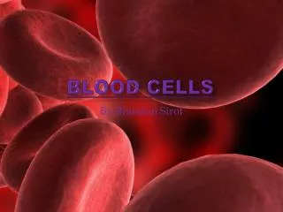
Erythrocytes (RBCs) • Biconcave disc • Folding increases surface area (30% more surface area) • Plasma membrane contains spectrin • Give erythrocytes their flexibility • Anucleate, no centrioles, no organelles • End result - no cell division • No mitochondria means they generate ATP anaerobically • Prevents consumption of O2 being transported • Filled with hemoglobin (Hb) - 97% of cell contents • Hb functions in gas transport • Hb + O2 HbO2 (oxyhemoglobin) • Most numerous of the formed elements • Females: 4.3–5.2 million cells/cubic millimeter • Males: 5.2–5.8 million cells/cubic millimeter
Erythrocytes (RBCs) Figure 17.3
Erythrocyte Function • Erythrocytes are dedicated to respiratory gas transport • Hemoglobin reversibly binds with oxygen and most oxygen in the blood is bound to hemoglobin • Composition of hemoglobin • A protein called globin • made up of two alpha and two beta chains • A heme molecule • Each heme group bears an atom of iron, which can bind to one oxygenmolecule • Each hemoglobin molecule thus can transport four molecules of oxygen
Structure of Hemoglobin Figure 17.4
Hemoglobin • Oxyhemoglobin – hemoglobin bound to oxygen • Oxygen loading takes place in the lungs • Deoxyhemoglobin – hemoglobin after oxygen diffuses into tissues (reduced Hb) • Carbaminohemoglobin – hemoglobin bound to carbon dioxide • Carbon dioxide loading takes place in the tissues
Fate and Destruction of Erythrocytes • The life span of an erythrocyte is 100–120 days • Travels about 750 miles in that time (LA to Albuquerque) • Old erythrocytes become rigid and fragile, and their hemoglobin begins to degenerate • Dying erythrocytes are engulfed by macrophages • Heme and globin are separated • Iron is removed from the heme and salvaged for reuse • Stored as hemosiderin or ferritinin tissues • Transported in plasma by beta-globulins as transferrin
Fate and Destruction of Erythrocytes • Heme is degraded to a yellow pigment called bilirubin • Liver secretes bilirubin into the intestines as bile • Intestines metabolize bilirubin into urobilinogen • Urobilinogen leaves the body in feces, in a pigment called stercobilin • Globin is metabolized into amino acids which are then released into the circulation
Laboratory Assessment of Blood Cells • Complete Blood Count (CBC) includes • White Blood Cell Count (WBC) • Red Blood Cell Count (RBC) • Percentage of white cells that are neutrophils, eosinophis or basophils (white cell differential count) • Amount of hemoglobin • Hematocrit • Percent of blood volume occupied by red blood cells
Erythrocyte Disorders • Polycythemia • Abnormal excess of erythrocytes • Increases viscosity, decreases flow rate of blood • Anemia • Abnormally low hemoglobin in blood • Caused by decreased numbers of RBC’s, decreased amount of hemoglobin in RBC’s, or both
ANEMIA • Hemoglobin level falls below normal range: • 14-18 g/dl for males and 12-16 g/dl for females • Signs and symptoms of hypoxia • Pallor, weakness, lethargy, and exercise intolerance • May affect cardiac rhythms and cause hepatic necrosis
Red Blood Cell Indices • Mean Corpuscular Volume (MCV) • Average size of a RBC • Mean Cell Hemoglobin (MCH) • Average amount of hemoglobin per RBC • Mean Corpuscular Hemoglobin Concentration (MCHC) • Average concentration of hemoglobin in all RBCs
Red Cell Indices Used to Diagnose Disease • Macrocytic • Red Blood Cells may be too large • Microcytic • Red Blood Cells may be too small • Normocytic • Red Blood Cells are normal size • Normochromic • Normal amount of hemoglobin • Hypochromic • Too little hemoglobin