Bright Field Images of Stem Cell Culture from Primary Isolation to 48 Hours
10 likes | 132 Vues
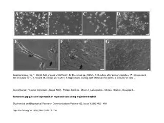
This figure illustrates bright field images of stem cells (SM) at four time points: 1, 2, 18, and 48 hours post-primary isolation. Each image captures the cellular morphology and recovery of cells during these intervals, providing insights into the growth and behavior of stem cells in cultured environments. Such observations are crucial for understanding cell dynamics and their applications in engineered tissues. The study by Sureshkumar Perumal Srinivasan et al. discusses enhanced gap junction expression in myoblast-containing engineered tissues.

Bright Field Images of Stem Cell Culture from Primary Isolation to 48 Hours
E N D
Presentation Transcript
Supplementary Fig. 1 Bright field images of SM from 1 to 48<ce:hsp sp="0.25"/> h of culture after primary isolation. (A–D) represent SM in culture for 1, 2, 18 and 48<ce:hsp sp="0.25"/> h respectively. During each of these time points, a recovery of cells ... Sureshkumar Perumal Srinivasan , Klaus Neef , Philipp Treskes , Oliver J. Liakopoulos , Christof Stamm , Douglas B.... Enhanced gap junction expression in myoblast-containing engineered tissue Biochemical and Biophysical Research Communications Volume 422, Issue 3 2012 462 - 468 http://dx.doi.org/10.1016/j.bbrc.2012.05.016