Download
1 / 32
320 likes | 507 Vues
Structural Basis of the DNA binding Domain of the p53 tumor suppressor protein. May 25, 2006 MOLECULAR DIAGNOSTICS Dr. Sandra Sharp. Jake Leon Department of Biology and Microbiology California State University Los Angeles 5151 State University Drive Los Angeles, CA 90032.

E N D
Structural Basis of the DNA binding Domain of the p53 tumor suppressor protein May 25, 2006 MOLECULAR DIAGNOSTICS Dr. Sandra Sharp Jake Leon Department of Biology and Microbiology California State University Los Angeles 5151 State University Drive Los Angeles, CA 90032
Normal Cells are able to prevent cancer by activating a natural defense mechanism • Cancer: • DNA damage • DNA damage activates gene expression • Genes code proteins • Proteins participate in response to DNA damage • p53 acts as a transcription factor that transactivates • genes to stop tumors by causing apoptosis, repairing DNA, or • by preventing cell proliferation.
Role of p53 in tumor suppression p53 knockout mice develop tumors spontaneously (Donehower et al., 1992) p53 binds to specific sequences on the DNA (Bargonetti et al., 1991; El-Deiry et al., 1992) p53 activates transcription of genes (Levine, 1997; Giaccia and Kastan, 1998) In human cancers, p53 is frequently observed to show mutations that inhibit its ability to bind to DNA
Outline of Next Series of Slides • Normal p53 in the cell when there is no detectable DNA damage • Normal p53 in response to DNA damage • 2 slides – repairable damage • Phopshorylation of p53 blocking its degradation and • Causing p53 to function as a transcription factor to stop the cell cycle • 2 slides – irreparable damage • Phosphorylation of p53 blocking its degradation and • Causing p53 to function as a transcription factor to induce apoptosis
Latent p53 p53 Half-life: ~20 min Nucleus This normally rapid turnover prevents normal cells from entering into cell cycle arrest or undergoing apoptosis MDM2 Murine Double Minute 2 26 S Proteosome Momand et al., 2005
ATP ATM ATM Ataxia Telangiectasia Mutagenesis ADP S15 p53 CHK2 Checkpoint Kinase 2 ADP S20 ATP MDM2
p21 GADD45 RNA polymerase p53 p53 p53 p53 TAF31 TAF70
ATP DNA-PK DNA-dependent protein kinase ADP S15 p53
These genes participate in the activation of APOPTOSIS Bax NOXA PUMA KILLER/DR5 Fas/Apo1 RNA polymerase p53 p53 p53 p53 TAF31 TAF70
Apoptosis Induction by p53 • P53 also responds to unrepaired DNA damage by inducing expression of genes that trigger apoptosis (programmed cell death) of the injured cell. • This ultimately leads to cell death. • DNA-PKc (catalytic subunit) is part of the enzymatic machinery for • VDJ rearrangement • Non-homologous end joining
Bax Apoptosis – in response to irreparable DNA damage Note the role of tumor suppressor p53.
What is p53? • A protein of ~53 kilodaltons • A nuclear phosphoprotein
DNA viruses and their oncogenes. • E6 is produced by Human Papilloma Virus (HPV) and can contribute to cervical cancer. • E1b is produced by Adenovirus. • Human cells are permissive for adenovirus. • Causes the common cold. • Adenovirus transforms rodent (non-permissive) cells • Human virus JC is similar to SV40 and may be associated with certain cancers, but a causative role has yet to be confirmed. • JC virus T antigen causes tumors in nude mice. • All these proteins are products of “early” genes in their viral replication cycles. • A productive infection of these viruses leads to lysis of the host cell. A non-productive infection allows the cells to live.
What is p53? • Transcriptional regulator • Binds to 12 bp recognition sequence in the promoters (regulatory regions) of the genes it regulates • Activates transcription by interacting with RNA polymerase complex
What is p53? • Acts as a tetramer • Individual molecules associate at tetramerization region • Oligomerization of mutated p53 with wt p53 inactive p53 complex
What is p53? • Detection of damaged DNA by p53 causes p53 to be stabilized and accumulate in the cell. • DNA damage activates the kinase ATM, which phosphorylates p53. • When damaged DNA is not present, p53 is turned over rapidly and does not accumulate because • the protein MDM2 binds to the transcription-activation region of p53 and targets p53 for degradation by a proteosome. (TAD) Note: MDM2 binds when the TAD is LESS phosphorylated.
p53 as a transcriptional regulator • If DNA damage is detected by binding of DNA fragments to the non-specific DNA binding region of p53, p53 stops DNA synthesis until the damage is repaired. • If DNA damage is detected, then • p53 is phosphorylated by a protein known as ATM • MDM2 is released from being bound to the transcriptional activation domain of p53 and • p53 is able to act as a transcriptional activator and turn on genes for • cyclin dependent kinase inhibitor p21, which • stops or prevents DNA synthesis • DNA repair • Example: GADD45 • If DNA damage is extensive and can not be repaired, p53 induces genes for apoptosis (programmed cell death).
p53 as a transcriptional regulator • p53 activates the gene for MDM2 • MDM2 • targets p53 for degradation and prevents inappropriate build up • prevents transcriptional activation by p53 • So, it’s a negative feedback loop! • p53 also turns expression of some genes off.
How does p53 inhibit DNA synthesis? Let’s work backwards. • E2F transcription factor turns on transcription of genes for DNA synthesis. • E2F can’t turn on genes if it is bound to Rb1, a tumor suppressor. • Rb1 can’t bind E2F if it is heavily phosphorylated. • Rb1 is phosphorylated by cyclin-dependent kinases (CDKs).
How does p53 inhibit DNA synthesis? • Cylin dependent kinases can be inhibited by cyclin dependent kinase inhibitors (CDKIs). If CDKs are inhibited • Rb1 won’t be phosphorylated • E2F will be bound by Rb1 • DNA synthesis genes will not be transcribed • And remember . . . . P53 induces expression of CDKI p21, a cyclin dependent kinase inhibitor! • Check out the next slide for a visual of these pathways.
This magnification of mutations in the DNA binding region of p53 gives more information regarding how the mutation affects p53. Note particularly that some mutations cause p53 to be misfolded (denatured) and others do not.
Have you figured it out? For our assay, the samples are cell extracts from two mouse cell lines, BC3H1 and C2C12. One line is wild type for p53; one is mutant. One accumulates detectable levels of p53; one doesn’t. Based on this lecture and your assay results, have you figured out which cell line does what? Have you thought about why? There is one explanation confirmed in the literature and at least one additional plausible contribution to what you observe. (Hint: P53 is not accumulating in either of these cell lines in response to DNA damage. DNA damage is a temporary condition which is repaired immediately. If it is not repaired, the cell soon dies as a result of apoptosis. Mutations may be the result of incorrect repair of DNA damage, but they are no longer considered damage because they are perfectly base-paired.)
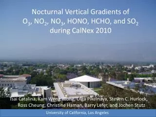
Zn p53 DBD Folding Cho, Y., Gorina, S., Jeffrey, P.D. and Pavletich, N.P. (1994) Science 265: 346-355
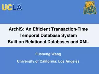
Crystal Structure of p53 DBD-DNA Complex PDB file 1tsr Cho, Y., Gorina, S., Jeffrey, P.D. and Pavletich, N.P. (1994) Science 265: 346-355
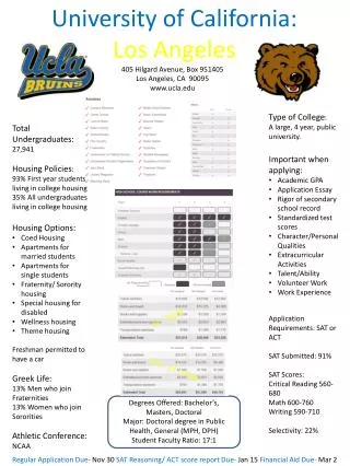
Crystal Structure of p53 DBD-DNA Complex PDB file 1tsr Cho, Y., Gorina, S., Jeffrey, P.D. and Pavletich, N.P. (1994) Science 265: 346-355
Crystal Structure of p53 DBD-DNA Complex PDB file 1tsr Cho, Y., Gorina, S., Jeffrey, P.D. and Pavletich, N.P. (1994) Science 265: 346-355
American Cancer Society: American Cancer Society: www. cancer.org • Bargonetti, J., Friedman, P., Kern, S., Vogelstein, B. and Prives, C. A. (1991) Cell65, 1083-1091 • Bertram, J.S. (2001) Mol. Aspects. Med. 21, 167-223 • Buzek, J., Latonen, L., Kurki, S., Peltonen, K. and Laiho, M. (2002) Nucleic Acids Res 30, 2340-2348 • Chehab, N. H., Malikzay, A., Appel, M. and Halazonetis, T. D. (2000) Gene Dev.14, 278-288. • Chehab, N. H., Malikzay, A., Stavridi, E. S. and Halazonetis, T. D. (1999) Proc. Natl. Acad. Sci.U S A.96, 13777-13782. • Chene, P. (1999) Biochem Biophys Res Commun263, 1-5 • Cho, Y., Gorina, S., Jeffrey, P. D. and Pavletich, N. P. (1994) Crystal structure of a p53 tumor suppressor-DNA complex: understanding tumorigenic mutations. Science 265:346-355,. • Delphin, C., Cahen, P., Lawrence J. J, and Baudier, J. (1994) Eur. J. Biochem.223, 683-692 • Donehower, L. A., Harvey, M., Slagle, B. L., McArthur, M. J., Montgomery, C. A., Butel, J. S. and Bradley, A. (1992) Nature356, 215-221 • El-Deiry, W. S., Kinzler, S. E., Pietenpol, J. A., Kinzler, K.W., Vogelstein B. (1992) Nat Genet1, 45 • Furuta, S., Ortiz, F., Sun, X., Wu, H., Mason, A., Momand, J. (2002) Biochem. J.365, 639-648. • Gaskel, S. (1997) Journal of Mass Spectrometry32, 677-688 • Giaccia, A. J. and Kastan, M. B. (1998) Genes Dev. 12, 2973-2983 • Hainaut, P. and Milner, J. (1993) Cancer Res.53, 4469-4473,. • Hainaut, P., Rolley, N., Davies, M. and Milner, J. (1995) Oncogene 10, 27-32 • Hirao, A., Kong, Y.Y., Matsuoka, S., Wakeham, A., Ruland, J., Yoshida, H., Liu, D., Elledge, S.J., and Mak, T.W. (2000) Science 287, 1824-1827 • Jayaraman, L., Murthy, K. G., Zhu, C., Curran, T., Xanthudakis, S., Prives, C. (1997) Genes Dev11, 558-570 • Kaeser, M., Iggo, R. (2002) PNAS99, 95-100. • Kim, S. T., Lim, D. S., Canman, C. E. and Kastan, M. B. (1999) J. Biol. Chem. 274, 37538-43 • Klein, C., Planker, E., Diercks, T., Kessler, H., Kunkele, K., Lang, K., Hansen, S., Schwaiger, M. (2001) The Journal of Biological Chemistry 276, 49020-49027. • Lee, S., Yang, K., Kwon, J., Lee, C., Jeong, W., Rhee, S., Reversible Inactivation of the Tumor Suppressor PTEN by H2O2. The Journal of Biological Chemistry 274, 20336-20342. • Levine, A. J. (1990) Virology177, 419-426. • Levine, A. J. (1997) Cell88, 323-331 • Levine, A. J., Momand, J. and Finlay, C. A. (1991) Nature351, 453-456 • Makmura, L., Hamman, M., Areopagita, A., Furuta, S., Muñoz, A. and Momand, J. (2001) Antioxid RedoxSignal3, 1105-1118 • Margalioth, E. J., Schenker, J. and Chevion, M. (1983) Cancer52, 868-872 • Mary, J., Vougier, S., Picot, C., Perichon, M., Petropoulos, I., Friguet, B. (2004) Experimental Gerontology 39, 1117-1123 • Matsuoka, S., Rotman, G., Ogawa, A., Shiloh, Y., Tamai, K., and Elledge, S.J. (2000) Proc. Natl. Acad. Sci. U S A. 97, 10389-394 • McLure, K., Lee, P., (1998) The EMBO Journal17, 3342-3350. • Momand, J., Wu, H., Dasgupta, G. (2000) Gene 242, 15-29 • Narayanan, V., Fitch, C., Levenson, C. (2001). Journal Nutrition 131, 1427-1432. • Oren, M. (1999) The Journal of Biological Chemistry 274, 36031-36034 • Peng, Y., Chen, L., Li, C., Lu, W., Agrawal, S., Chen, J. (2001) The Journal of Biological Chemistry 276, 6874-6878. • Protein Data Bank (PDB): http//:www.rcsb.org/pdb • Rainwater, R., Parks, D., Anderson, M. E., Tegtmeyer, P. and Mann K. (1995) Mol. Cell. Biol.15, 3892-3903 • Siliciano, J., Canman, C., Taya, Y., Sakaguchi, K., Appella E., Kastan, M. (1997) Genes &Development 11, 3471-3481 • Smith, M., Ford, J., Hollander, M., Bortnick, R., Amundson, S., Seo, Y., Deng, C., Hanawalt, P., Fornace, A. (2000) Molecular and Cellular Biology20, 3705-3714 • Smith, M.L., Ford, J. M., Hollander, M. C., Bortnick, R.A., Amundson, S. A., Seo Y. R., Deng, C.X., Hanawalt, P. C., and Fornace A. J. Jr. (2000). Mol. Cell. Biol. 20, 3705-3714 • Standing, K. (2003) Current Opinion in Structural Biology13, 595-601 • Sun, X., Vinci, C., Makmura, L., Han, S., Tran, D., Nguyen, J., Hamann, M., Grazziani, S., Sheppard, S., Gutova, M., Zhou, F., Thomas, J. and Momand, J. (2003) Antioxidants & Redox Signaling5, 655-665. • Wang, P., Sait, F., Winter, G. (2001) Oncogene20, 2318-2324. • Wang, S., Guo, M., Ouyang, H., Li, Z., Cordon-Cardo, C., Kurimasa, A., Chen, D. J., Fuks, Z., Ling, C.C., and Li, G.C. (2000) Proc. Natl. Acad. Sci.U S A.97, 1584-1588 • Wu, H. H., Sherman, M., Yuan, Y. C. and Momand, J. (1999) Gene Ther. Mol.Biol.4, 119-132 • Wu, H., Thomas, J. and Momand, J. (2000) Biochem. J.351, 87-93. • Xiao, G., Chicas, A., Oliver, M., Taya, Y., Tyagi, S., Kramer, F.R., and Bargonetti, J. (2000) Cancer Res. 60, 1711-1719 • Yang, H., Wen, Y., Chen, C., Lozano, G., Lee, M. (2003) Mol. Cell. Biol.23, 7096-7107.