Download
1 / 8
140 likes | 1.02k Vues
Ultrasound Case: Myositis Ossificans of Rectus Femoris Muscle. Diagnosis: Myositis Ossificans of Rectus Femoris muscle History: Female patient, soccer player, sustained blow to the thigh, with development of palpable mass.

E N D
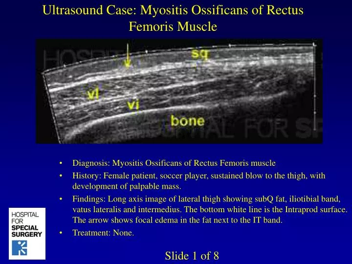
Ultrasound Case: Myositis Ossificans of Rectus Femoris Muscle • Diagnosis: Myositis Ossificans of Rectus Femoris muscle • History: Female patient, soccer player, sustained blow to the thigh, with development of palpable mass. • Findings: Long axis image of lateral thigh showing subQ fat, iliotibial band, vatus lateralis and intermedius. The bottom white line is the Intraprod surface. The arrow shows focal edema in the fat next to the IT band. • Treatment: None. Slide 1 of 8
Ultrasound Case: Myositis Ossificans of Rectus Femoris Muscle • Diagnosis: Myositis Ossificans of Rectus Femoris muscle • History: Female patient, soccer player, sustained blow to the thigh, with development of palpable mass. • Findings: Transverse image over the lateral extensor surface showing focal edema in the fat (arrow) just over the iliotibial band (IT). This appears like a small white ellipse in the adjacent fat. • Treatment: None. Slide 2 of 8
Ultrasound Case: Myositis Ossificans of Rectus Femoris Muscle • Diagnosis: Myositis Ossificans of Rectus Femoris muscle • History: Female patient, soccer player, sustained blow to the thigh, with development of palpable mass. • Findings: Long axis view of the rectus femoris muscle shows an organizing hematoma (arrow) which is calcifying. The calcification produces an acoustic shadow (sh) which appears below the hematoma. It’s dark because no sound gets beyond the calcification. The rectus muscle to the right is bright, due to edema.. • Treatment: None. Slide 3 of 8
Ultrasound Case: Myositis Ossificans of Rectus Femoris Muscle • Diagnosis: Myositis Ossificans of Rectus Femoris muscle • History: Female patient, soccer player, sustained blow to the thigh, with development of palpable mass. • Findings: Color siescape image shows neovascularity at site of organizing hematoma. The shadow (sh) within the muscle arises due to calcification. Numerous prominent intramuscular vessels are apparent. • Treatment: None. Slide 4 of 8
Ultrasound Case: Myositis Ossificans of Rectus Femoris Muscle • Diagnosis: Myositis Ossificans of Rectus Femoris muscle • History: Female patient, soccer player, sustained blow to the thigh, with development of palpable mass. • Findings: Transverse image over the thigh showing the extensor muscles in cross-section. Here we see the rectus, vatus lateralis and intermedius. • Treatment: None. Slide 5 of 8
Ultrasound Case: Myositis Ossificans of Rectus Femoris Muscle • Diagnosis: Myositis Ossificans of Rectus Femoris muscle • History: Female patient, soccer player, sustained blow to the thigh, with development of palpable mass. • Findings: Long axis image over the medial thigh in a child with a large soft to palpation mass. Extended field of view image shows this to be confined to the fat, containing multiple dark tubular structures. The mass mildly deforms but doesn’t directly invade the deep muscles. (see next slide). • Treatment: None. Slide 6 of 8
Ultrasound Case: Myositis Ossificans of Rectus Femoris Muscle • Diagnosis: Myositis Ossificans of Rectus Femoris muscle • History: Female patient, soccer player, sustained blow to the thigh, with development of palpable mass. • Findings: Long axis view at different plane shows the mass to have large cystic spaces. No flow demonstrated within the mass on Doppler imaging. The imaging features suggest this to be a lymphangioma. • Treatment: None. Slide 7 of 8
Ultrasound Case: Myositis Ossificans of Rectus Femoris Muscle • Diagnosis: Myositis Ossificans of Rectus Femoris muscle • History: Female patient, soccer player, sustained blow to the thigh, with development of palpable mass. • Findings: Long axis view of infraspinatus muscle and tendon to insertion on greater tuberosity. There is mild inhomogeneity (*) in the tendon, which should normally be bright. • Treatment: None. Slide 8 of 8