Download
1 / 42
480 likes | 1.48k Vues
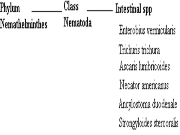
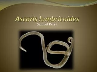
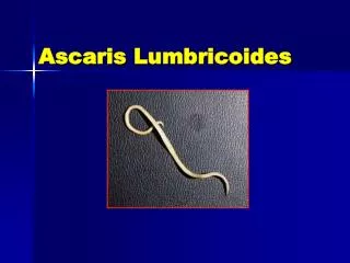
NEMATODES Ascaris Lumbricoides. NEMATODES (Round Worms). Ascaris lumbricoides (roundworm), Trichinella spiralis (trichinosis), Trichuris trichiura (whipworm), Enterobius vermicularis (pinworm), Strongyloides stercoralis (Cochin-china diarrhea),

E N D
NEMATODES (Round Worms) • Ascaris lumbricoides (roundworm), • Trichinella spiralis (trichinosis), • Trichuris trichiura (whipworm), • Enterobius vermicularis (pinworm), • Strongyloides stercoralis (Cochin-china diarrhea), • Ancylostoma duodenale and Necator americanes (hookworms)
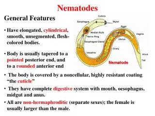
Nematodes or roundworms are among the most abundant animals on earth - over 500,000 species have been described. • Majority of nematodes are free-living in every conceivable habitat.
Characteristics of the Phylum Nematoda Reproductive system consists of tubular organs lying in the pseudocoelom. MALE nematodes are generally smaller in size.
Characteristics of the Phylum Nematoda FEMALE nematodes are larger in size. The female reproductive organs are doubled.
Characteristics of the Phylum Nematoda Nematode development is similar in all nematodes. Consists of 4 larval (=juvenile) stages between the egg and adult. Each stage is separated by a molt of the cuticle. M1 M2 M3 M4 Egg L1 L2 L3 L4 Adult Larval stages may be passed within the egg, free-living in soil, parasitic in an intermediate host, or parasitic in definitive host.
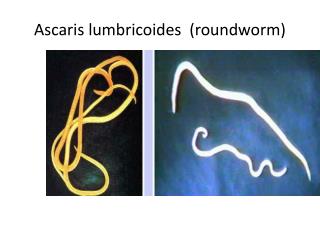
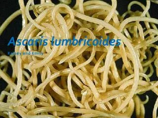
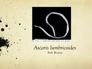
Ascaris Lumbricoides • Ascaris lumbricoides , commonsaying “round worm of man”, is the largest of the intestinal nematodes parasitizing humans. • It is the most common worm found in human. • It is worldwide in distribution and most prevalent through out the tropics, sub-tropics and more prevalent in the countryside than in the city.
Ascaris lumbricoides.The incidence is over 1500 million infections annually. Of these cases, about 210 million are symptomatic. In some rural settings with poor sanitation, perhaps half the children of 2-12 years have ascariasis.Then many of them will also have trichuriasis and various of other chronic illnesses.
I. Morphology • Adult: The adults are cylindrical in shape, creamy-white or pinkish in color. • The female averages 20-35cm in length, the largest 49cm. • The male is smaller, averaging 15-31cm in length and distinctly more slender than the female. • The typical curled tail with a pair sickle like copulatory spines. On the tip of the head there are three lips, arranged as a Chinese word “品 ”.
They have a complete digestive tract. • Reproductive organs are tubular. male has a single reproductive tubule. The female has two reproductive tubules and the vulva is ventrally located at the posterior part of the anterior 1/3 of the body.
Ascaris lumbricoides Female Male
Ascaris lumbricoides cont. Female gut oviduct genital pore uterus vagina
Ascaris lumbricoides cont. Male sperm duct seminal vesicle testes gut
Lumbricus A Worm’s Death
Lumbricus cont. clitellum
Lumbricus cont. seminal vesicle spermathecae crop septa gizzard
Lumbricus cont. Intestine with chlorogogue cells septum nephridium ventral nerve cord
The lips of Ascaris lumbricoides The three lips are seen at the anterior end. The margin of each lip is lined with minute teeth which are not visible at this magnification.
Egg: There are three kinds of the eggs. They are fertilized eggs, unfertilized eggs and decorticated eggs. We usually describe an egg in 5 aspects: size, color, shape, shell and content.
1. Fertilized eggs: broad oval in shape, brown in color, an average size 60× 45µm. The shell is thicker and consists of ascaroside, chitinous layer, fertilizing membrane and mammillated albuminous coat stained brown by bile. The content is a fertilized ovum. There is a new-moon(crescent) shaped clear space at the each end inside the shell.
Fertilized Ascaris Egg A fertilized Ascaris egg, still at the unicellular stage, as they are when passed in stool.
2.Unfertilized egg: Longer and slender than a fertilized egg. The chitinous layer and albuminous coat are thinner than those of the fertilized eggs without ascaroside and fertilizing membrane. The content is made of many refractable granules various in size.
3. Decorticated eggs: Both fertilized and unfertilized eggs sometimes may lack their outer albuminous coats and are colorless.
II Life Cycle • 1.Site of inhabitation: small intestine 2. Infective stage: embryonated eggs 3. Route of infection: by mouth 4. No intermediate and reservoir hosts 5. Life span of the adult: about 1 year
This worm lives in the lumen of small intestine, feeding on the intestinal contents, where the fertilized female lays eggs. • An adult female can produce approximately 240,000 eggs per day, which are passed in feces. • When passed, the eggs are unsegmented and require outside development of about three weeks until a motile embryo is formed within the egg.
After the ingestion of embryonated eggs in contaminated food or drink or from contaminated fingers, host digestive juices acts on the egg shell and liberate the larva into the small intestine. • These larvae penetrate the intestinal mucosa and enter lymphatics and mesenteric vessels. • They are carried by circulation to the liver, right heart and finally to the lungs where they penetrate the capillaries into the alveoli in which they molt twice and stay for 10-14days and then they are carried,
or migrate, up the bronchioles, bronchi, and trachea to the epiglottis. • When swallowed, the larvae pass down into the small intestine where they develop into adults. • The time from the ingestion of embryonated eggs to oviposition by the females is about 60-75 days. • The adult worms live for about one year. The ascarid life cycle is as the following diagram.
Ascaris lumbricoidesworms have a reputation for wandering, and often do so if the body they are in-the host-is ill or taking certain medications. • Adult roundworms sometimes spontaneously exit the host through the anus, mouth, or nose. • They are found in the bathtub, toilet bowl, in diapers, or even on the pillow upon waking.
III. Pathogenesis There are two phase in ascariasis: 1. The blood-lung migration phase of the larvae: During the migration through the lungs, the larvae may cause a pneumonia. The symptoms of the pneumonia are low fever, cough, blood-tinged sputum, asthma. Large numbers of worms may give rise to allergic symptoms. Eosionophilia is generally present. These clinical manifestation is also called Loeffler’s syndrome.
2. The intestinal phase of the adults. The presence of a few adult worms in the lumen of the small intestine usually produces no symptoms, but may give rise to vague abdominal pains or intermittent colic, especially in children. A heavy worm burden can result in malnutrition. More serious manifestations have been observed. Wandering adults may block the appendical lumen or the common bile duct and even perforate the intestinal wall.
Thus complications of ascariasis, such as intestinal obstruction, appendicitis, biliary ascariasis, perforation of the intestine, cholecystitis, pancreatitis and peritonitis, etc., may occur, in which biliary ascariasis is the most common complication.
Iii. Diagnosis • The symptoms and signs are for reference only. The confirmative diagnosis depends on the recovery and identification of the worm or its egg. 1. Ascaris pneumonitis: examination of sputum for Ascaris larvae is sometimes successful. 2. Intestinal ascariasis: feces are examined for the ascaris eggs. (1) direct fecal film: it is simple and effective. The eggs are easily found using this way due to a large number of the female oviposition, approximately 240,000 eggs per worm per day. So this method is the first choice.
(2) brine-floatation method: (3) recovery of adult worms: when adults or adolescents are found in feces or vomit and tissues and organs from the human infected with ascarids , the diagnosis may be defined.
V. Epidemiology • World wide distribution, very common in China, especially in the countryside. Factors favoring the spread of the transmission: 1.Simple life cycle. 2. Enormous egg production ( 240,000 eggs/ day/ female ).
3. These eggs are highly resistant to ordinary disinfectants( due to the ascroside). The eggs may remain viable for several years. 4. Social customs and living habits. 5. Disposal of feces is unsuitable.
VI. Prevention and Treatment • 1.Treatment to ascariasis:Mebendazole, Albendazole and Levamizole are effective. • 2.Sanitary disposal of feces. • 3.Hygienic habits such as cleaning of hands before meals. • 4.Health education.