Download
1 / 29
350 likes | 1.03k Vues
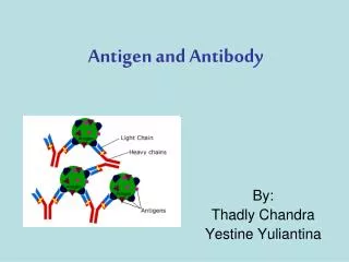
Immunodiagnosis Antigen antibody reactions. -Antigen antibody reactions in vitro are known as serological reactions . -They can be used for detection and quantification of either antigens or antibodies(Diagnosis of infectious diseases, autoimmune diseases, determination of blood groups).

E N D
ImmunodiagnosisAntigen antibody reactions -Antigen antibody reactions in vitro are known as serological reactions. -They can be used for detection and quantification of either antigens or antibodies(Diagnosis of infectious diseases, autoimmune diseases, determination of blood groups). - Antigens & antibodies combine with each other specifically and in observable manner. - Measurement of antigen and antibody is in terms of *mass (example mg) *Units or titre. -The antibody titreis the highest dilution of the serum which shows an observable reaction with the antigen in a particular test. - Antigens also can be titrated against sera.
Antigen antibody reactions(serological tests) • Agglutination reactions • Precipitation reactions • Toxin-antitoxin neutralization test • Virus neutralization test • Complement fixation test • Immunofluorescence test • Radioimmuno assay • Enzyme- linked immunosorbent assay(ELISA • Immunoelectrobloteg., westrenblot • Immunochromatographictest.
Agglutination reactions The antigen is in form of particles(Suspension suspended antigen)e.g. RBCs, bacteria, latex particles coated with antigens. • When a particulate antigen is mixed with its specific antibody the particles are clumped or agglutinated. • If one of the reactants(Ag or Ab) is known, the reaction can be used for identification of the other.
Types of agglutination reactions I- Direct agglutination: A-Slide agglutination test: On a clean slide, place a suspension of unknown culture of an organism, then add to it a drop of known antiserum(AB), and mix well→ clumping(Agglutination) →Positive →No clumping → Negative This test is commonly used in blood grouping. negative positive
B-Tube agglutination test • Is a quantitative test, used to determine the amount of antibodies in the patient serum (titer). • Done by mixing serial dilutions of the patient serum (1/20, 1/40, 1/80, etc.) with fixed amount of a known bacterial suspension, suspected to be the cause of the disease. The tubes are incubated over night, then examined for presence of clumping at the bottom of the tubes with clear supernatant fluid • The last tube showing visible agglutination will reflect the serum antibody titer. -The antibody titreis the highest dilution of the serum which shows an observable agglutination reaction with the antigen. NB: Paired serum samples separated by 1-2 weeks are recommended in serodiagnosis. A rising titre of 2 folds or more is diagnostic. e.g. Widal test in diagnosis of enteric fever, in diagnosis of Malta fever, ………
2- Antiglobulin agglutination test (Coomb's test) • Used to detect Rhincompatibility which causes erythroblastosis fetalis. • Also used to detect incomplete Abs in autoimmune hemolytic anemia. • Most anti-RH antibodies are IgGincomplete antibodies which can only coat the Rh positive RBCs, but can not cause agglutination. 1- Indirect Comb's test: detects anti-RhAbs or autoantibodiesin the serum. Mother’s serum is mixed with washed Rh+ve RBCs blood group (O)→incubate for 1-2 hr→ Centrifuge→ wash the deposit →add antihumanglobulon→again incubate→Agglutination(+Ve). 2- Direct Comb's test: detects incomplete Abs coating the RBCs of Thenewborn. The antihuman globulon is added directly to a washed suspension of newborn RBCS.
3- Passive agglutination test Agglutination reaction in which inert particles as latex or RBCs are coated with various Ags or Abs. These particles are aggregated in the presence of specific Abs or Ags respectively. e.g. 1- Pregnancy test. 2- Rheumatoid factor (RF). 3- C-reactive protein. 4- ASOT
4- Coagglutination(CoA) This test is based on that, Staph.aureus is rich in protein A. Staph.aureusis coated with Abs and mixed with unknown Ag→Agglutination(+Ve). 5- Virus haemagglutination inhibition(HAI) Serum(?Abs) + known Ag(Virus)→incubate for 1 hr, then add washed RBCs→ Incubate for ¼ hr +ve =No HA(No agglutination for RBCs) -ve =HA(agglutination for RBCs)
Prozone & zone of equivalence & zone of antigen excess (postzone) • Precipitation will occur more rapidly and abundantly when antigens & antibodies are present in optimal or equivalent proportions. • When antibody excess or antigen excess the precipitation is absent or weak. • Prozone– antibody excess, many antibodies coat all antigen sites- results in false negative • Postzone– antigen excess, antibody coats antigen but cannot get lattice formation, results in false negative
Precipitation • The antigen is soluble in solution, Ag-Ab reaction results in formation of a precipitate. • Methods of PPT reactions: A-Tube precipitation test(Ring test): this simplest type of precipitation test consists of layering of antiserum in narrow tube (preferably capillary tube). A precipitate forms at junction of the two liquids.
B- PPT in agar gel (Immunodiffusion): Diffusion of Ag and Ab is allowed to occur in agar gel. 1- Double diffusion (Ouchterlony): Wells are punched in agar. • The Ag is placed in one well and the Ab in another. Both will diffuse towards each other and where they meet at optimal proportions, PPT bands (Lines)will form. • 3 types of lines:a) line of identity,b) line of non-identity, c) line of partial identity
2-Radial (Single)immunodiffusion • The antibody is incorporated in the agar gel and the antigen is placed in a well cut in the agar. • The antigen reacts with the antibody to give precipitate ring around the well. • The diameter of the ring is proportional to the concentration of each antigen • Estimations can be done by comparing with known concentration of antigen. Uses of radial immunodiffusion • Estimation of plasma proteins • Immunoglobulins (IgG, IgM,) • Complement proteins.etc.,
3- Precipitation in agar with an electric field: A- Immunoelectrophoresis: - A serum sample is placed in a well in agar on a slide. - An electric current is passed through the agar, the serum proteins will move in the electric field according to their molecular weight and charge. - A trough is cut into the agar and filled with antiserum antibody. • As the Ags and Abs diffuse towards each other, PPT are will be formed. • This permits serum proteins to be characterized in terms of presence or absence or unusual patterns as in human myloma proteins.
B- Western Blot: • Antigen, or mixture of antigens is first separated electrophoretically in gel. • The separated materials are then blotted (transferred) onto nitrocellulose sheet, to which antigen bind strongly. • Antibody is then applied and will bind to its specific antigen. • To visualize the result of test, the antibody is labeled with enzyme or radioactive substance (direct test), or a labeled anti-immunoglobulin is added. e.g. WB test used to confirm diagnosis of HIV infection.
Complement fixation test(CFT) Serum(? Abs)+ known Ag+C→Incubate for 1 hour, then add indicator system(Sheep RBCs+Ani-sheep RBCs).→Incubate for 1 hour +ve = No lysis -ve= lysis Uses: Diagnosis of: Syphilis, Viral Infections, Gonorrhoea,….
Immunofluoresence 1-Direct: For detection of Ag Specimen(? Ag) fixed on a slide + Fluoresceinlabeled Abs.→ Incubate for 1 hour then wash and examine under flu.microscope +ve = green fluoresence -ve = absence of fluoresence 2-Indirect: For detection of Abs Serum(? Abs)+Known Ag,→ incubate for 1 hr, and then wash. Add specific labeled anti-human γ globulin,→ incubate for 1 hr, and then wash. Examine under flu.microscope. Uses: Syphilis, viral infection,………..
Virus neutralization test Serum(? Abs) + Ag(virus)→incubate for 1 hr. → Inoculate this mixture to tissue culture→ Incubate for several days. +ve = No CPE -ve = Presence of CPE
Enzyme linked immunosorbent assay(ELISA) 1- Double Abtechnique (sandwich technique) :For detection of Ag Known Ab is fixed to the solid phase + Specimen(? Ag),→incubate for 1 hr and then wash. Add specific labelledAbs,→incubatefor 1 hr and then wash. Add substrate , →measure the colour+ve = Colour -ve = No colour 2- Indirect: For detection of Abs: Serum(?Abs)+known Ag fixed to the solid phase, → incubate for 1 hr and then wash. Add labelled anti-human γ-globulin,→incubate for 1 hr and then wash. Add substrate →measure the colour +ve = Colour -ve = No colour
ELISA PRINCIPLE OF ELISA
Radioimmunoassay • In radioimmunoassay instead of fluorescent dyes radioactive material is used • Radioactivity is measured after the reaction in gamma counter. • Used for detection of thyroid hormones T3 & T4 & HBs Ag.
Toxin-antitoxin neutralization test Toxin + Serum(Antitoxin)→ ½ hr→RBCs suspension(Blood group O)→ ½ hr Result: No hemolysis→+ve Hemolysis → -Ve e.g.Antistreptolysin O titre (ASOT) used for diagnosis of rheumatic fever
Immunochromatographic test(IC) • It is produced in card with immunological reagents(either Ag or Ab) fixed on the card membrane.The specimen to be tested is applied to the well(S) • Result: • 2 pink bands (C & T)→+Ve • 1 pink band(Only in c) →-Ve • Uses: For HIV, HBV, HCV, Pregnancy,H.pylori, AFP…..