Download
1 / 24
240 likes | 666 Vues
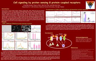
G Protein Linked Receptors. Introduction. G-protein-linked receptors are the largest family of cell-surface receptors. More than 100 members have already been defined in mammals.

E N D
Introduction • G-protein-linked receptors are the largest family of cell-surface receptors. • More than 100 members have already been defined in mammals. • G-protein-linked receptors mediate the cellular responses to an enormous diversity of signaling molecules, including hormones, neurotransmitters, and local mediators, which are as varied in structure as they are in function: the list includes proteins and small peptides, as well as amino acid and fatty acid derivatives. • The same ligand can activate many different family members. • At least 9 distinct G-protein-linked receptors are activated by adrenaline, for example, another 5 or more by acetylcholine, and at least 15 by serotonin. • Despite the chemical and functional diversity of the signaling molecules that bind to them, all of the G-Protein-linked, G-protein-linked receptors whose amino acid sequences are known from DNA sequencing studies have a similar structure and are almost certainly evolutionarily related. • They consist of a single polypeptide chain that threads back and forth across the lipid bilayer seven times
A schematic drawing of a G-protein-linked receptor. Receptors that bind protein ligands have a large extracellular ligand-binding domain formed by the part of the polypeptidemchain shown in light green. Receptors for small ligands such as adrenaline have small extracellular domains, and the ligand-binding site is usually deep within the plane of the membrane, formed by amino acids from several of the transmembrane segments. The parts of the intracellular domains that are mainly responsible for binding to trimeric G proteins are shown in orange,while those that become phosphorylated during receptor esensitization are shown in red
Trimeric G Proteins Relay the Intracellular Signal from G protein- linked Receptors • The trimeric GTP-binding proteins (G proteins) that functionally couple these receptors to their target enzymes or ion channels in the plasma membrane are structurally distinct from the single chain GTP-binding proteins (called monomeric GTP-binding proteins or monomericGTPases) that help relay intracellular signals and regulate vesicular traffic and many other processes in eucaryotic cells. • Both classes of GTP-binding proteins, however, are GTPases and function as molecular switches that can flip between two states: active, when GTP is bound, and inactive, when GDP is bound. • When an extracellular ligand binds to a G-protein-linked receptor, the receptor changes its conformation and switches on the trimeric G proteins that associate with it by causing them to eject their GDP and replace it with GTP. • The switch is turned off when the G protein hydrolyzes its own bound GTP, converting it back to GDP. • But before that occurs, the active protein has an opportunity to diffuse away from the receptor and deliver its message for a prolonged period to its downstream target.
Most G-protein-linked receptors activate a chain of events that alters the concentration of one or more small intracellular signaling molecules. • These small molecules, often referred to as intracellular mediators (also called intracellular messengers or second messengers), in turn pass the signal on by altering the behavior of selected cellular proteins. • Two of the most widely used intracellular mediators are • cyclic AMP (cAMP) and • Ca2+: changes in their concentrations are stimulated by distinct pathways in most animal cells, and most G-protein-linked receptors regulate one or the other of them
Two major pathways by which G-protein-linked cell-surface receptors generate small intracellular mediators. In both cases the binding of an extracellular ligand alters the conformation of the cytoplasmic domain of the receptor, causing it to bind to a G protein that activates (or inactivates) a plasma membrane enzyme. In the cyclic AMP (cAMP) pathway the enzyme directly produces cyclic AMP. In the Ca2+ pathway the enzyme produces a soluble mediator that releases Ca2+ from the endoplasmic reticulum. Like other small intracellular mediators, both cyclic AMP and Ca2+ relay the signal by acting as allosteric effectors: they bind to specific proteins in the cell, altering their conformation and thereby their activity.
Some Receptors Increase Intracellular Cyclic AMP byActivating Adenylyl Cyclase via a Stimulatory G Protein (Gs) • Cyclic AMP was first identified as an intracellular mediator of hormone action in 1959 and has since been found to act as an intracellular signaling molecule in all procaryotic and animal cells that have been studied. • For cyclic AMP to function as an intracellular mediator, its intracellular concentration (normally <10-7 M) must be able to change up or down in response to extracellular signals: upon hormonal stimulation, cyclic AMP levels can change fivefold in seconds. • Such responsiveness requires that rapid synthesis of the molecule be balanced by rapid breakdown or removal. • Cyclic AMP is synthesized from ATP by a plasma-membrane-bound enzyme adenylylcyclase, and it is rapidly and continuously destroyed by one or more cyclic AMP phosphodiesterases, which hydrolyze cyclic AMP to adenosine 5‘- monophosphate (5'-AMP)
Cyclic AMP. It is shown as a formula, a ball-and-stick model, and as a space-filling model. (C, H, N, O, and P indicate carbon, hydrogen, nitrogen, oxygen, and phosphorus atoms, respectively.)
The synthesis and degradation of cyclic AMP (cAMP). A pyrophosphatase makes the synthesis of cyclic AMP an irreversible reaction by hydrolyzing the released pyrophosphate
Many extracellular signaling molecules work by controlling cyclic AMP levels, and they do so by altering the activity of adenylylcyclase rather than the activity of phosphodiesterase. • Just as the same steroid hormone produces different effects in different targe cells, so different target cells respond very differently to external signals that change intracellular cyclic AMP levels. • All ligands that activate adenylylcyclase in a given type of target cell, however, usually produce the same effect: • at least four hormones activate adenylylcyclase in fat cells, for example, and all of them stimulate the breakdown of triglyceride (the storage form of fat) to fatty acids
Adenylyl cyclase. In vertebrates the enzyme usually contains about 1100 amino acid residues and is thought to have two clusters of six transmembrane segments separating two similar cytoplasmic catalytic domains. There are at least six types of this form of adenylyl cyclase in mammals (types I-VI). All of them are stimulated by Gs, but type I, which is found mainly in the brain, is also stimulated by complexes of Ca2+ bound to the Ca2+-binding protein calmodulin
The different receptors for these hormones activate a common pool of adenylylcyclase molecules, to which they are coupled by a trimeric G protein. • Because this G protein is involved in enzyme activation, it is called stimulatory G protein (Gs). • Individuals who are genetically deficient in Gs show decreased responses to certain hormones and, consequently, have metabolic abnormalities, abnormal bone development, and are mentally retarded. • The best-studied examples of receptors coupled to the activation of adenylylcyclase are the b-adrenergic receptors, which mediate some of the actions of adrenaline and noradrenaline
Trimeric G Proteins Are Thought to Disassemble When Activated • A trimeric G protein is composed of three different polypeptide chains, called a, b, and g. • The Gs a chain(as) binds and hydrolyzes GTP and activates adenylylcyclase. • The Gs b chain and g chain form a tight complex (bg), which anchors Gs to the cytoplasmic face of the plasma membrane, at least partly by a lipid chain (a prenyl group) that is covalently attached to the g subunit. • In its inactive form Gs exists as a trimer with GDP bound to as. • When stimulated by binding to a ligand activated receptor, as exchanges its GDP for GTP. • This is thought to cause as to dissociate from b g, allowing as to bind instead to an adenylylcyclase molecule, which it activates to produce cyclic AMP.
If cells are to be able to respond rapidly to changes in the concentration of an extracellular signaling molecule, the activation of adenylylcyclase must be reversed quickly once the signaling ligand dissociates from its receptor. • This ability to respond rapidly to change is assured because the lifetime of the active form of as is short: the GTPase activity of as is stimulated when as binds to adenylylcyclase, so that the bound GTP is hydrolyzed to GDP, rendering both as and the adenylylcyclase inactive. • The as then reassociates with bg to re-form an inactive Gs molecule
Assignment: Role of G protein in Cholera
Some Receptors Decrease Cyclic AMP by Inhibiting Adenylyl Cyclase via an Inhibitory Trimeric G Protein (Gi) • The same signaling molecule can either increase or decrease the intracellular concentration of cyclic AMP depending on the type of receptor to which it binds. • When adrenaline binds to b- adrenergic receptors, for example, it activates adenylylcyclase, whereas when it binds to a2- adrenergic receptors, it inhibits the enzyme. • The difference reflects the type of G proteins that couple these receptors to the cyclase. • While the b-adrenergic receptors are functionally coupled to adenylylcyclase by Gs, the a2-adrenergic receptors are coupled to this enzyme by an inhibitory G protein (Gi). • Gi can contain the same b-g complex as Gs, but it has a different a subunit (ai). • When activated, a2-adrenergic receptors bind to Gi, causing ai to bind GTP and dissociate from the bg complex. • Both the released ai and bg are thought to contribute to the inhibition of adenylylcyclase. • ai inhibits the cyclase, probably indirectly, whereas bg may inhibit cyclic AMP synthesis in two ways - directly, by binding to the cyclase itself, and indirectly, by binding to any free as subunits in the same cell, thereby preventing them from activating cyclase molecules.
Assignment: Role of G protein in Pertusis
Cyclic-AMP-dependent Protein Kinase (A-Kinase) Mediates the Effects of Cyclic AMP • Cyclic AMP exerts its effects in animal cells mainly by activating the enzyme cyclic-AMP dependent protein kinase (A-kinase), which catalyzes the transfer of the terminal phosphate group from ATP to specific serines or threonines of selected proteins. • A-kinase is found in all animal cells and is thought to account for all of the effects of cyclic AMP in most of these cells. • In the inactive state A-kinase consists of a complex of two catalytic subunits and two regulatory subunits that bind cyclic AMP. • The binding of cyclic AMP alters the conformation of the regulatory subunits, causing them to dissociate from the complex. • The released catalytic subunits are thereby activated to phosphorylate specific substrate protein molecules
The activation of cyclic-AMP-dependent protein kinase (A-kinase). The binding of cyclic AMP to the regulatory subunits induces a conformational change, causing these subunits to dissociate from the complex, thereby activating the catalytic subunits. Each regulatory subunit has two cyclic-AMP-binding sites, and the release of the catalytic subunits requires the binding of more than two cyclic AMP molecules to the tetramer. This greatly sharpens the response of the kinase to changes in cyclic AMP concentration. There are at least two types of A kinase in most mammalian cells: type I is mainly in the cytosol, whereas type II is bound via its regulatory subunit to the plasma membrane, nuclear membrane, and microtubules. In both cases, however, once the catalytic subunits are freed and active, they can migrate into the nucleus (where they can phosphorylate gene regulatory proteins), while the regulatory subunits remain in the cytoplasm.
Glycogen metabolism in skeletal muscle cells • Glycogen is the major storage form of glucose, and both its synthesis and degradation in skeletal muscle cells are regulated by adrenaline. • When an animal is frightened or otherwise stressed, for example, the adrenal gland secretes adrenaline into the blood, "alerting" various tissues in the body. • Among other effects, the circulating adrenaline induces muscle cells to break down glycogen to glucose 1-phosphate and at the same time to stop synthesizing glycogen. • The glucose is then oxidized by glycolysis to provide ATP for sustained muscle contraction. • In this way adrenaline prepares the muscle cells for anticipated strenuous activity. • Adrenaline acts by binding to b-adrenergic receptors on the muscle cell surface, thereby causing an increase in the level of cyclic AMP in the cytosol. • The cyclic AMP activates A-kinase, which phosphorylates two other enzymes.
The first, phosphorylasekinase, which was the first protein kinase to be discovered (in 1956), phos-phorylates in turn the enzyme glycogen phosphorylase, thereby activating the phosphorylase to release glucose residues from the glycogen molecule • The second enzyme phosphorylated by activated A-kinase is glycogen synthase, which performs the final step in glycogen synthesis from glucose. • This phosphorylation inhibits the enzyme's activity, thereby shutting off glycogen synthesis. • By means of this cascade of interactions, an increase in cyclic AMP levels both stimulates glycogen breakdown and inhibits glycogen synthesis, thus maximizing the amount of glucose available to the cell.
The stimulation of glycogen breakdown by cyclic AMP in skeletal muscle cells. The binding of cyclic AMP to A-kinase activates this enzyme to phosphorylate and thereby activate phosphorylase kinase, which in turn phosphorylates and activates glycogen phosphorylase, the enzyme that breaks down glycogen. The A-kinase also directly and indirectly increases the phosphorylation of glycogen synthase, which inhibits the enzyme, thereby shutting off glycogen synthesis.
In some animal cells an increase in cyclic AMP activates the transcription of specific genes. • In cells that secrete the peptide hormone somatostatin, for example, cyclic AMP turns on the gene that encodes this hormone. • The regulatory region of the somatostatin gene contains a short DNA sequence, called the cyclic AMP response element (CRE), that is also found in the regulatory region of other genes that are activated by cyclic AMP. • This sequence is recognized by a specific gene regulatory protein called CRE-binding (CREB) protein. • When CREB is phosphorylated by A kinase on a single serine residue, it is activated to turn on the transcription of these genes; the phosphorylation stimulates the transcriptional activity of CREB without affecting its DNA-binding properties. • If this serine residue is mutated, CREB is inactivated and no longer stimulates gene transcription in response to a rise in cyclic AMP levels.