Infrared Spectroscopy
Infrared Spectroscopy. Electromagnetic Radiation. The propagation of electromagnetic radiation in a vacuum is constant for all regions of the spectrum (= velocity of light): c = × 1 Å = 10 –10 m 1 nm = 10 –9 m 1 m = 10 –6 m.


Infrared Spectroscopy
E N D
Presentation Transcript
Electromagnetic Radiation The propagation of electromagnetic radiation in a vacuum is constant for all regions of the spectrum (= velocity of light): c = × 1 Å = 10 –10 m 1 nm = 10 –9 m 1 m = 10 –6 m Another unit commonly used is the wavenumber, which is linear with energy: Work by Einstein, Planck and Bohr indicated that electromagnetic radiation can be regarded as a stream of particles or quanta, for which the energy is given by the Bohr equation:
IR Spectroscopy • Introduction • The entire electromagnetic spectrum is used by chemists: Frequency, nin Hz ~1019 ~1017 ~1015 ~1013 ~1010 ~105 Wavelength, l ~.0001 nm ~0.01 nm 10 nm 1000 nm 0.01 cm 100 m Energy (kcal/mol) > 300 300-30 300-30 ~10-4 ~10-6 g-rays X-rays UV IR Microwave Radio Visible
An IR Spectrum • A plot of % transmittance vs vibrational frequency in wavenumbers (cm-1) λ= wavelength υ= frequency c = speed of light in a vacuum
Wavenumbers • The higher the wavenumber, the shorter the wavelength.
An IR Spectrum from http://www.cem.msu.edu/~reusch/VirtualText/Spectrpy/InfraRed/infrared.htm
An IR Spectrum • The wavelength of IR radiation is in the 2.5-25 micron range (compare to visible light in the 400-700 nm range). • The frequencies of IR radiation are more conveniently expressed by a wavenumber (cycles per cm), than by (cycles per 3 x 1010 cm).
Characteristic Vibrational Frequencies of Bonds • Bonds are not rigid but behave like a spring with a mass at either end. • Obey Hooke’s Law: F = -kx • This gives rise to a characteristic frequency for the vibration:
Characteristic Vibrational Frequencies of Bonds • Characteristic frequency for the vibration: • The frequency is affected by • the masses of the atoms in the bond • the strength of the bond
Characteristic Vibrational Frequencies of Bonds • The lower the mass, the higher the vibrational frequency. • Stretching frequencies for bonds to carbon: • C-H > C-C > C-N > C-O
Characteristic Vibrational Frequencies of Bonds • The stronger the bond, the higher the vibrational frequency. • Stretching frequencies • C≡C > C=C > C-C • C≡N > C=N > C-N • C≡O > C=O > C-O • C(sp)-H > C(sp2)-H > C(sp3)-H
Number of Vibrational Frequencies in a Molecule • There are 3n-6 possible vibrational modes in a nonlinear molecule with no symmetry • Symmetry reduces the number of possible vibrational modes. • Water has 3 possible vibrational modes. • Formaldehyde has 6.
The Fingerprint Region is Unique to the Molecule • In addition, the vibration of a particular bond in a molecule affects the whole molecule. • The various harmonics of a bond vibration can combine and lead to a number of combinational bands. • The intensity of these bands is usually 1/100 the intensity of the main vibrational absorptions. • These make up the “fingerprint region.” (occur at <1250 cm-1)
Intensity of IR Absorptions • In order for a vibration mode to absorb in the infrared, the vibrational motion must cause a change in the dipole moment of the bond. • The intensity of the IR “peaks” is proportional to the change in dipole moment that a bond undergoes during a vibration. • C=O bonds absorb strongly. • C=C bonds generally absorb much less.
How to Analyze an IR Spectrum • Pay the most attention to the strongest absorptions: • -C=O • -OH • -NH2 • -C≡N • -NO2 • Pay more attention to the peaks to the left of the fingerprint region (>1250 cm-1).
How to Analyze an IR Spectrum • Pay the most attention to the strongest absorptions. • Pay more attention to the peaks to the left of the fingerprint region (>1250 cm-1). • Note the absence of certain peaks. • Be wary of O-H peaks, water is a common contaminant.
Characteristic IR Wavenumbers *The peak is broad when H bonding is extensive. Otherwise, the peak can be sharp.
How to Analyze an IR Spectrum • Look for what’s there and what’s not there. • C-H absorption • The wavenumber will tell you sp3(C-C), sp2(C=C), sp (C≡C) and perhaps aldehyde. • Carbonyl (C=O) absorption • Its presence means the compound is an aldehyde, ketone, carboxylic acid, ester, amide, anhydride or acyl halide. • Its absence means the compound cannot be any of the carbonyl-containing compounds.
How to Analyze an IR Spectrum • O-H or N-H absorption • This indicates either an alcohol, N-H containing amine or amide, or carboxylic acid. • C≡C and C≡N absorptions • Be careful: internal triple bonds often do not show up in IR spectra. • C=C absorption • Can indicate whether compound is alkene or aromatic.
How to Analyze an IR Spectrum • N-O of NO2 absorption • This is a distinctive, strong doublet that it pays to know (1515-1560 & 1345-1385 cm-1). • Read the scale for the value of the wavenumbers (be able to interpolate), or • Read the wavenumbers in the table provided.
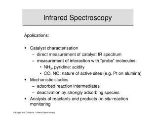
IR Spectroscopy • Introduction • The IR Spectroscopic Process • The quantum mechanical energy levels observed in IR spectroscopy are those of molecular vibration • We perceive this vibration as heat • When we say a covalent bond between two atoms is of a certain length, we are citing an average because the bond behaves as if it were a vibrating spring connecting the two atoms • For a simple diatomic molecule, this model is easy to visualize:
Infrared region LIMIT OF RED LIGHT: 800 nm, 0.8 m, 12500 cm-1 NEAR INFRARED: 0.8 -2.5 m, 12500 - 4000 cm-1 MID INFRARED: 2.5 - 50 m, 4000 - 200 cm-1 FAR INFRARED:50 - 1000 m, 200 - 10 cm-1 Divisions arise because of different optical materials and instrumentation.
Infrared radiation λ = 2.5 to 50 μm υ = 4000 to 200 cm-1 These frequencies match the frequencies of covalent bond stretching and bending vibrations. Infrared spectroscopy can be used to find out about covalent bonds in molecules. IR is used to tell: 1. what type of bonds are present 2. some structural information
Molecular spectra • There are three basic types of optical spectra that we can observe for molecules: • Electronic or vibronic spectra (UV-visible-near IR) • (transitions between a specific vibrational and rotational level of one electronic state and a vibrational and rotational level of another electronic state) • Vibrational or vibrational-rotational spectra (IR region) • (transitions from the rotational levels of one vibrational level to the rotational levels of another vibrational level in the same electronic state) • Rotational spectra (microwave region) • (transitions between rotational levels of the same vibrational level of the same electronic state)
Vibrational spectra (II): Anharmonic oscillator model The actual potential energy of vibrations fits the parabolic function fairly well only near the equilibrium internuclear distance. The Morse potential function more closely resembles the potential energy of vibrations in a molecule for all internuclear distances-anharmonic oscillator model. Fig. 12-1
EVIB= ( En+1 – En) =h× osc • The energy difference between the transition from n to n+1 corresponds to the energy of the absorbed light quantum • The difference between two adjacent energy levels gets smaller with increasing n until dissociation of the molecule occurs (Dissociation energy ED ) Note: Weaker transitions called “overtones” are sometimes observed. These correspond to =2 or 3, and their frequencies are less than two or three times the fundamental frequency (=1) because of anharmonicity. Typical energy spacings for vibrational levels are on the order of 10-20 J. from the Bolzmann distribution, it can be shown that at room temperature typically 1% or less of the molecules are in excited states in the absence of external radiation. Thus most absorption transitions observed at room temperature are from the =0 to the =1 level.
IR Spectroscopy • Introduction • The IR Spectroscopic Process • There are two types of bond vibration: • Stretch – Vibration or oscillation along the line of the bond • Bend – Vibration or oscillation not along the line of the bond H H H H C C C C C C H H H H H H C C C C H H asymmetric symmetric scissor rock twist wag in plane out of plane
Infrared Spectroscopy • The IR Spectroscopic Process • As a covalent bond oscillates – due to the oscillation of the dipole of the molecule – a varying electromagnetic field is produced • The greater the dipole moment change through the vibration, the more intense the EM field that is generated
Infrared Spectroscopy IR beam from spectrometer “coupled” wave EM oscillating wave from bond vibration • The IR Spectroscopic Process • When a wave of infrared light encounters this oscillating EM field generated by the oscillating dipole of the same frequency, the two waves couple, and IR light is absorbed • The coupled wave now vibrates with twice the amplitude
Infrared Spectroscopy • The IR Spectrum • Each stretching and bending vibration occurs with a characteristic frequency as the atoms and charges involved are different for different bonds The y-axis on an IR spectrum is in units of % transmittance In regions where the EM field of an osc. bond interacts with IR light of the same n – transmittance is low (light is absorbed) In regions where no osc. bond is interacting with IR light, transmittance nears 100%
IR source è sample è prism è detector graph of % transmission vs. frequency => IR spectrum 100 %T 0 4000 3000 2000 1500 1000 500 v (cm-1)
IR spectra of ALKANES C—H bond “saturated” (sp3) 2850-2960 cm-1 + 1350-1470 cm-1 -CH2- + 1430-1470 -CH3 + “ and 1375 -CH(CH3)2 + “ and 1370, 1385 -C(CH3)3 + “ and 1370(s), 1395 (m)
n-pentane 2850-2960 cm-1 sat’d C-H 3000 cm-1 1470 &1375 cm-1 CH3CH2CH2CH2CH3
n-hexane CH3CH2CH2CH2CH2CH3
cyclohexane no 1375 cm-1 no –CH3
IR of ALKENES =C—H bond, “unsaturated” vinyl (sp2) 3020-3080 cm-1 + 675-1000 RCH=CH2 + 910-920 & 990-1000 R2C=CH2 + 880-900 cis-RCH=CHR + 675-730 (v) trans-RCH=CHR + 965-975 C=C bond 1640-1680 cm-1 (v)
1-decene unsat’d C-H 3020-3080 cm-1 910-920 & 990-1000 RCH=CH2 C=C 1640-1680
4-methyl-1-pentene 910-920 & 990-1000 RCH=CH2
2-methyl-1-butene 880-900 R2C=CH2
2,3-dimethyl-1-butene 880-900 R2C=CH2
IR spectra BENZENEs =C—H bond, “unsaturated” “aryl” (sp2) 3000-3100 cm-1 + 690-840 mono-substituted + 690-710, 730-770 ortho-disubstituted + 735-770 meta-disubstituted + 690-710, 750-810(m) para-disubstituted + 810-840(m) C=C bond 1500, 1600 cm-1
ethylbenzene 3000-3100 cm-1 Unsat’d C-H 1500 & 1600 Benzene ring 690-710, 730-770 mono-
o-xylene 735-770 ortho
p-xylene 810-840(m) para