Download
1 / 104
1.08k likes | 1.41k Vues
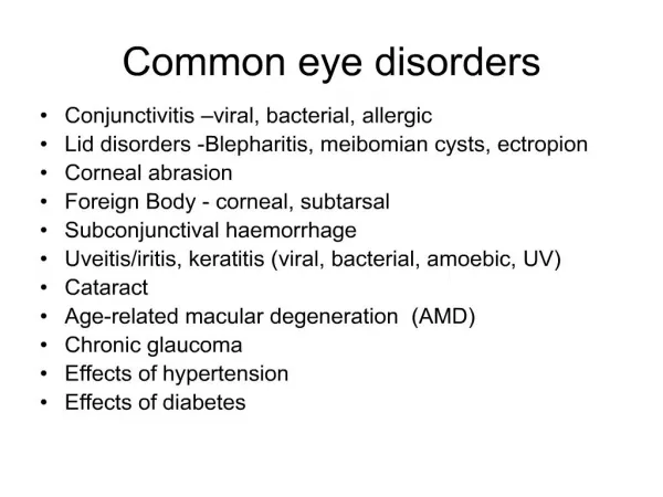
Disorders of the Eye. Objectives. Describe common disorders of the eye Describe inflammatory conditions of the lid, conjunctiva, and cornea Describe the clinical manifestations of common disorders and inflammatory conditions of the eye. Objectives.

E N D
Objectives • Describe common disorders of the eye • Describe inflammatory conditions of the lid, conjunctiva, and cornea • Describe the clinical manifestations of common disorders and inflammatory conditions of the eye
Objectives • Discuss nursing assessment and nursing interventions for eye conditions • Discuss Diagnostic Tests, medical management, and prognosis for eye conditions • Discuss patient teaching needs
Blindness and Near-Blindness • Etiology/pathophysiology • Loss of visual acuity that ranges from partial to total loss of sight (includes no light perception) • Functional blindness = some light perception but no usable vision
Blindness or Near-Blindness • Can be: Congenital or Acquired • Legal blindness refers to a maximum visual acuity of: • 20/200 with corrective eyewear (normal 20/20) • Visual field range less than 20 degrees (normal 180)
Blindness or Near-Blindness • Congenital blindness results from various birth defects • Acquired blindness in adults: • Diabetic retinopathy • Glaucoma • Cataracts • Retinal degeneration • Acute trauma
Blindness or Near-Blindness • Acquired Blindness • Clinical manifestations/assessment • Diplopia • Pain • Floaters and light flashes • Pruritus; burning of the eyes • Loss of peripheral vision • Halos (rainbow colors seen around lights) • A sense of orbital pressure • Bulging of the eyes • Difference in appearance of an eye structure • Emotional symptoms
Blindness or Near-Blindness • Medical Management • Corrective eyeware • Assistive devices such as canes, guide dogs, magnifying systems, and telescopic lenses • Surgical correction
Blindness or Near-Blindness • Assessment • Note patient complaint of blurred vision • Determine onset, severity and duration of symptoms • Note any factors that relieve symptoms • Observe for squinting and rubbing the eyes • Note compensation measures (eg. Use of magnifying glass) • Impaired self-care
Effect of Visual Impairment Mild losses may require only some adaptations Serious losses affect independence, mobility, employment, and interpersonal relationships People grieve for the lost function just as they might grieve after the death of a loved one Factors that affect a person’s response to this loss include personality, usual coping style, effect of vision loss on the person’s life, and the circumstances of the loss
Care of the Visually Impaired Patient • Be aware of visually impaired person’s thoughts and feelings about handicaps/disabilities • Assume that people with visual impairments can be independent and productive • The person needs help with some tasks but should be treated as an adult • The extent of vision loss determines the types of assistance needed
Care of the Visually Impaired Patient • Interventions • Comprehensive approach • Educate, Assist, Counsel • Prevention of complications • Nursing Diagnosis (AEB, R/T) • Risk for Injury r/t environmental hazards • Disturbed Sensory Perception • Ineffective Coping • Self-Care Deficit • Ineffective Therapeutic Regimen Management
Refraction • Light rays bend (refract) as they pass through the lens • Enables light from the environment to focus on the retina • Refractory errors indicate that the light is not hitting the correct spot on the retina
Refractory Errors • Etiology/pathophysiology of common refractory errors • Astigmatism – defect in the curvature of the eyeball surface • Strabismus—inability of the eyes to focus in one direction; cross-eyed • Myopia-nearsightedness; eyeball is elongated • Hyperopia- farsightedness; eyeball is too short
Astigmatism • Clinical Manifestations: • Blurring of vision = primary manifestation • Assessment: not c/o eye discomfort; mainly blurring, difficulty focusing • Diagnostic Tests: opthalmoscopy, retinoscopy, visual acuity test, and refraction tests.
Astigmatism • Medical Management: • corrective eyewear • surgical correction • Nursing Intervention and Patient Teaching: • Assistance with ADLs prn • Make sure eyewear is clean • Safety when eyewear is not worn
Strabismus • Strabismus: inability of the eye to focus in the same direction [“cross-eyed”] • Clinical Manifestations: eyeball position is not symmetrical d/t neurological or muscular dysfunction • Assessment: c/o difficulty in following an object; same is observed • Diagnostic tests, Medical Management, and Nursing Interventions: same as for Astigmatism
Myopia • The medical term for nearsightedness • The lens is situated too far from the retina • Light rays come together to focus in front of the retina instead of on the retina • The retina only receives a fuzzy image People with myopia have difficulty seeing distant images clearly • Snellen test useful • New glasses needed approximately every 2 years • Refractory surgery: ages 20-60
Myopia • Medical Management • Keratorefractive Surgery • Surgery to alter the corneal curvature • Surgeon uses a special microsurgical knife or laser to open and replace a flap of corneal tissue • Photorefractive Keratectomy (PRK) • Laser is used to reshape the corneal surface • LASIK (Laser-in-situ Keratomileasis)
Myopia • Nursing Intervention and Patient Teaching • Preop – Instruct pt. to stop wearing hard contact lens 1-2 dayss before surgical evaluation • Eye drop routine
Myopia • Post Op • Eye patch post op • Can be up and around at home though should be encouraged to rest the first day • Assistance PRN • Medication Instruction: oral analgesics • Instruct s/sx complications: infection, when to contact MD • Reinforce need to keep MD follow-up appointments • Usually seen the next day; then at 1 week post-op; then monthly for 1 year
Hyperopia • Commonly known as farsightedness • The lens is too close to the retina • Light rays come together behind the retina producing a fuzzy image The hyperopic person sees clearly in the distance but has difficulty focusing on close objects • Convex corrective lenses needed • Diagnostic tests, medical management, Nursing interventions same as for Myopia. Pt. Teaching: care of corrective lenses or contact lenses
Amblyopia • Commonly referred to as “lazy eye” - an inaccurate label • Poor vision due to brain favoring one eye. The weaker eye tends to wander inward or outward even with correction • Common in children • Diagnosis confirmed when decreased visual acuity cannot be explained by an organic cause. • Treatment: corrective eyewear; eye patches to make the weaker eye work
Presbyopia • Inability to focus on close objects • Poor accommodation due to loss of elasticity of the ciliary muscles • Accommodation: adjustment of the lens for near and distant vision • Contraction or relaxation of the ciliary muscles, which causes the lens to change shape • It most often develops after age 40 • Corrective lenses are needed
Refractory Errors • Diagnostic tests • Opthalmoscopy • Retinoscopy • Visual acuity tests • Refraction tests
Blepharitis • Inflammation of eyelid along eyelid margin • Ulcerative Blepharitis: Caused by bacteria, most often by staphylococci • NonUlcerative Blepharitis: caused by psoriasis, seborrhea, or allergic response
Blepharitis • Symptoms: • Erythema of eyelid • Eyelid pain • Photophobia • Scales or crusts on the lid margins • Excessive tearing in nonulcerative type
Blepharitis • Assessment: • Ulcerative type: pt. c/o eye itching, lids adhering together during sleep • NonUlcerative: red eyes, patient rubs eyes, sensitivity to light, tear spillage
Blepharitis • Medical Management: • Antibiotic ointment (Erythromycin) • NOTE: Be certain that any medication applied to the eye is an ophthalmic preparation • Eyelids can be gently cleansed with baby shampoo solution
Nursing Interventions and Patient Teaching • Primary Objective prevention of spread of infection • Instruct on use of prescribed eye drops or ointment • Teach Handwashing, avoidance of irritating perfumes or smoke. The use of make-up should be avoided until all inflammation subsides – then use NEW make-up
Hordeolum • Commonly called a “stye” • Acute staphylococcal infection (abcess) of the eyelid margin that originates in a lash follicle or sebaceous gland of the eyelashes • Affected area of lid is red, swollen, and tender • Treatment - Apply warm, moist compresses several times a day 10-20 min • Repeated infections may be related to staphylococcal infections at some other location on the body • Physician may treat with ophthalmic antibiotics
Chalazion • Inflammation of the meibomian (sebum) glands in the eyelids (may become a cyst) • Swelling prevents fluid from leaving the glands, causing tenderness Treatment: • Warm compresses may bring some relief • Physician may order antibiotics if infection is present • Surgical removal of the gland necessary if condition persists
Conjunctivitis • An inflammation of the conjunctiva caused by: • Bacterial or viral infection • Allergy • Environmental factors • Commonly called pink eye
Conjunctivitis • Bacterial Conjunctivitis • Caused by streptococcal, staphlococcal, gonococcal, pneumococcal, chlamydial organisms • HIGHLY CONTAGIOUS! • HANDSare the most common transmitters of the bacteria • Infected people should practice good hand washing and should avoid sharing washcloths
Conjunctivitis • Symptoms: • Red conjunctiva, • Mild irritation (gritty feeling) • Edema of eyelid • Drainage overnight (crusty) • Diagnostic Test: conjunctiva scraped for bacteria and stained for microscopic exam
Nursing Interventions and Patient Teaching • Treat with warm compresses and topical antibiotics • Cleanse lid and lashes with NS • Warm compresses 2-4x/day • When allergies are present: cold compress to reduce edema • Eye irrigations • Gentamycin, Erythromycin, Tobramycin, Ciprofloxacin
Nursing Interventions and Patient Teaching • Instruct client to avoid contact with eyes or soiled material when infection present. Use individual washcloths • Wash hands before treatment, and when contact is made with eyes **WEAR GLOVES! • Avoid noxious fumes and smoke • Avoid using contact lenses during the inflammation period
Conjunctivitis • Viral conjunctivitis • Caused by: herpes simplex virus type 1, herpes zoster virus, or Chlamydia trachomatis • Characterized by redness and drainage • Round, raised white or gray areas on the conjunctiva • Infections caused by herpes simplex virus type 1 are treated with antiviral ointments (Acyclovir) or other topical medications
Keratitis • Etiology/pathophysiology • Inflammation of the cornea • Due to: Injury, irritants, allergies, viral infection, or diseases • Ulcers may form in the eye membrane layers scattered scarring of the corneal surface • Pneumococcus, staphylococcus, streptococcus, and pseudomonas are most common types of bacterial causes • Herpes simplex is most common viral cause