Skin Assessment
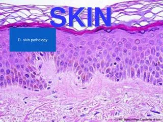
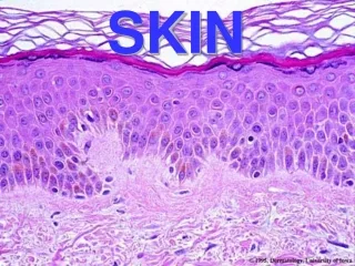
Skin Assessment. Skin Assessment. Skin is the largest organ in the body Skin is composed of Epidermis- outermost portion of a relatively uniform, thin but tough, composed of thickness stratum germinativum and stratum corneum a. color derived from three sources Brown- pigment melanin

Skin Assessment
E N D
Presentation Transcript
Skin Assessment • Skin is the largest organ in the body • Skin is composed of • Epidermis- outermost portion of a relatively uniform, thin but tough, composed of thickness stratum germinativum and stratum corneum • a. color derived from three sources • Brown- pigment melanin • Yellow-orange tones of pigment carotene • Red-purple tone in underlying vascular bed
2. Dermis- bulk of skin; the inner supportive layer consisting mostly of connective tissue or collagen is tough fibrous protein that enables skin to resist tearing and allows skin to stretch with movement. 3. Subcutaneous layer- adipose tissue made up of lobules of fat cells used for energy. It provides insulation for temperature control and aids in protection by its soft cushioning effect
Skin function • Protection/Barrier • Sensation • Temperature regulation • Identification • Communication • Wound Repair • Absorption/Excretion • Vitamin D
Assess Skin of Adults 1. Previous history of skin diseases 2. Skin Color- affected by genetic factors and physiological factors. -Variations of skin color • Cyanosis- blue tinge • Pallor- loss of rosy glow in skin, paleness • Erythema- redness of the skin, increase in climate temperature, inflammation, infection
Plethora- redness of skin caused by increase red blood cell • Ecchymosis- large diffuse areas usually black and blue , results of injuries • Petechiae- small pinpoint hemorrhages can denote some type of blood disorder • Jaundice- yellow staining of skin usually caused by bile pigments
3. Changes in mole • size, shape, tenderness, bleeding • check for abnormal characteristics of pigmented lesions. • Note any freckles and changes and any birthmarks (report any changes in size, itching, burning, bleeding of moles)
Abnormal characteristics of pigmented lesions: • ABCDE • Asymmetry of pigmented lesion -one that is not regularly round or oval • Border irregularity -notching, scalloping, ragged edges or poorly defined margins • Color variation -areas of brown, tan, black, blue, red, white or combination • Diameter greater than 6mm • Elevation and enlargement
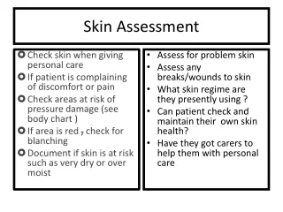
Texture- palpate note any marks or scaring skin should be smooth and firm • Temperature- symmetrically feel each part of the body, compare upper area with lower areas check for hypothermia and hyperthermia • Normal finding: warm • Changes: cool, cold, hot
Turgor-amount of elasticity in skin, grasp index finger pull it taut and quickly release- elastic skin immediately assumes in normal position, poor turgor suspended or tented; turgor shows hydration and nutrition • Moisture or dryness- check face, hands, axilla, skin folds; shows diaphoresis or dehydration • Are there any rashes or lesions; note color, elevation, pattern or shapes, size, location and distribution on body, any exudates
Is there any itching (purities) • What medication are you taking • Note mobility • Note any edema- accumulation of fluid in the intercellular spaces; to check for edema, imprint your thumbs firmly against the ankle malleolus or the tibia. If pressure leaves a dent in the skin “pitting” present 1+= mild pitting, slight indentation, no perceptible swelling of the leg 2+= moderate pitting, indentation subsides rapidly
3+= Deep pitting, indentation remains for a short time; leg looks swollen 4+=Very Deep pitting, indentation lasts a long time, leg is very swollen. • thickening uniform over body except thick over palms and soles of feet
Depress pretibial area & medial malleolus for 5 seconds Grade pitting edema 1+ to 4+ Assessing for Edema
Accessory structure of skin of adults • Hair- • inspect for color (comes from melanin) graying may begin at 3rd decade; • Texture maybe fine or thick; straight, curly, or kinky; • Quality maybe shinny or dull; • Distribution- coarse or elastic 2.Scalp- inspect for ticks or lice 3. Nails- Shape and Contour- curved or flat, edges smooth, rounded, clean; - Consistency- smooth, regular, nor brittle or splitting, thickness, firm - Color- translucent, pink nails base - inspect nail beds for clubbing
Capillary return or refill: normal = less than 3 seconds • used to evaluate the ability of the circulatory system to restore blood to the capillary system (perfusion). • Capillary refill is evaluated at the nail bed in a finger. • Place your thumb on the patient’s fingernail and gently compress. • Pressure forces blood from the capillaries. • Release the pressure and observe the fingernail. • As the capillaries refill, the nail bed returns to its normal deep pink color. • Capillary refill should be both prompt and pink. • Color in the nail bed should be restored within 2 seconds, about the time it takes to say "capillary refill."
Monitoring Skin Condition • Check color • Temperature • Abnormalities • Excessive dryness, moisture, itching, flaking • General texture of skin • Skin turgor • Edema • Cleanliness • Odor • Discoloration (ecchymosis, petechiae, purpura, erythema, altered pigmentation)
Vocabulary • Alopecia • Hirsutism • Clubbing of nail • Onycholysis
Benefits and Disadvantages • Subjective, no quantitative data • Requires experience/training • May not indicate subclinical damage • Surface conditions do not always correlate with conditions in the ☺ • Quick • Inexpensive • Can be done ‘on-site’ • Valid for all visible skin conditions
Practice • Go to this website for a tutorial on skin assessment • http://www.logicalimages.com:80/morphology/morphology3_content.html