Download
1 / 46
460 likes | 695 Vues
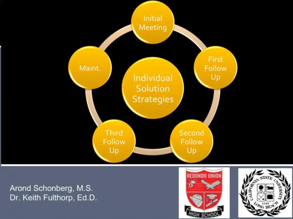
Dr. Rajan Modi M.S. F.M.A.S. THE BASICS OF VENOUS DISEASE: What You Should Know. Premier Laser Vein Clinic. An Introductory Lecture. 2. “ It is ironic that medical education does not cover three of the most common medical problems: back pain,. hemorrhoids, and varicose veins. ”.

E N D
Dr. RajanModiM.S. F.M.A.S. THE BASICS OF VENOUS DISEASE:What You Should Know. Premier Laser Vein Clinic An Introductory Lecture 2
“It is ironic that medical education does not cover three of the most common medical problems: back pain, hemorrhoids, and varicose veins.” P. Fujimura, MD Surgical Intern University of California School of Medicine
PHLEBOLOGY The medical specialty devoted to the diagnosis and treatment of patients with venous disorders
Anatomy and physiology of the venous system Deep venous system: the channel through which 90% of venous blood is pumped out of the legs Superficial venous system: the collecting system of veins Perforating veins: the conduits for blood to travel from the superficial to the deep veins Musculovenous pump: Contraction of foot and leg muscles pumps the blood through one-way valves up and out of the legs
Valve Failure Prolonged dilation damages the valves, so the blood flows in the wrong direction The resulting backpressure is termed “venous hypertension”, and this is what causes symptoms
IMPORTANCE OF CHRONIC VENOUS DISEASE Incidence increases with age and is more common in women with over 40% of women in their 50’s suffering from some sort of venous disorder
Etiology of Venous Insufficiency Inherited genetic predisposition Normal aging process Hormonal changes History of venous thrombosis Excess pressure on leg veins Standing Obesity Pregnancy Tumors Constipation Previous surgery
Venous Disease is a Hereditary Disorder The risk of developing varicose veins was: 89% if both parents had varicose veins 47% if one parent had varicose veins 20% if neither parent had varicose veins
Other Factors Chronic Venous Insufficiency Although some risk factors for venous disease such as age, family history of venous disease are immutable others can be modified, such as weight, physical activity, and cigarette smoking.
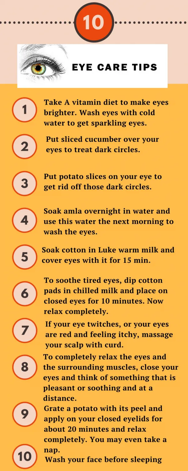
Presenting Symptoms of Chronic Venous Disease • Aching 17percent • Cramping-14 • Tired legs-12 • Swelling-12 Heaviness-12 Itching -6 Restless legs-6
THE SPECTRUM OF CHRONIC VENOUS DISEASE Superficial phlebitis telangiectasias venous ulceration 5 varicose veins lipodermatosclerosis
Venous Disease Classification CEAP What? How ? Where? Why? Clinical Etiologic Anatomic Pathologic
Venous Disease Classification Clinical C0 - No visible or palpable venous disease C1 - Telangectasias or reticular veins C2 - Varicose veins C3 - Edema C4 - Skin changes (a - pigmentation b - dematosclerosis) C5 - Healed ulcers C6 - Active ulcers
Venous Disease Classification Etiological C - congenital P - primary (venous failure without other cause) S - secondary (post-phlebitic syndrome)
Venous Disease Classification Anatomical S - superficial D - deep P - perforator
Venous Disease Classification Pathological R - reflux O - obstruction
Venous Disease Diagnosis Physical Exam Visible veins Swelling Ulceration Skin changes Standing venous reflux exam Duplex ultrasound with compression and release
Standing Venous Reflux Exam Establishes presence of reflux Deep and/or superficial systems Results dictate treatment option Superficial only Conservative or interventional Deep only Conservative Deep and superficial involvement Conservative Selective interventional
Venous Disease Treatment Options Conservative Elevation Compression Interventional Stripping Laser ablation • Radio frequency ablation Micro or stab phlebectomy • Sclerotherapy
Conservative Treatment Compression Therapy
Treatment of telangiectasias Sclerotherapy most effective Laser may be helpful Multiple treatments usually required Reduces symptoms in 85% of patients Improves quality of life
Surgical Treatment of Varicose Veins: Vein Stripping Vein stripping used to remove Great and Small saphenous veins
Conventional Treatment Crossectomie and Stripping ► Very invasive and traumatic procedure ► General anaesthesia is required ► Destroys the connective tissue ► Poor cosmetical result due to scarring ► Patients have to be hospitalised► Recovery time 2-3 weeks ► High Recurrence Rates
EVLT Less invasive technique: Endovenous Laser Treatment (EVLT). EVLT uses targeted laser energy to seal the vein shut
Why ELVeS™ - Advantages GSV treatment • Gentle, moderate and yet effective method of treatment • Less traumatic • Minimal discomfort • Quick and easy to perform • No scarring • No general anaesthesia required • Excellent aesthetic results • Recurrence rates are extremely low • Outpatient Procedure • A rapid return to normal activities ELVeS™by biolitec
EVLT Procedure A catheter is placed in the vein through a needle utilizing ultrasound guidance • The laser is passed through the catheter to the saphenofemoral junction which is confirmed by ultrasound
ELVeS™ - The Procedure for the GSV Map the Course of the Vein
ELVeS™ - The Procedure for the GSV Access to the Vein • Anaesthesia by local intradermal injection • Entry of the needle into the vein under Ultrasound Guidance
ELVeS™ - The Procedure for the GSV • After the puncture the guide wire is inserted into the vein • The entry needle is withdrawn and the introducer sheath and dilator are inserted over the guide wire and into the vein • The guide wire is then removed and the laser fibre is inserted into the introducer sheath and advanced along its length • The introduction of the laser fiber can be monitored by using the aiming beam ELVeS™by biolitec
ELVeS™ - The Procedure for the GSV • The fibre is advanced to a position 2-3 centimeters below the sapheno-femoral junction to preserve the integrity of the femoral vein from the first pulse of laser energy • The final position 2-3 cm below the sapheno femoral junction is controlled by ultrasound ELVeS™by biolitec
EVLT Advantages Relief of chronic pain symptoms Faster return to normal activity Improves venous stasis wound healing potential Decreases wound recurrence Minimal-to-no scarring Typically is covered by insurance
EVLT Post-op Care Dress leg with compression bandage Class II compression stocking on POD 2 Resume normal activities as tolerated Avoid hot baths Avoid vigorous gym workouts
EVLT Post-op Mild Bruising can occur along the leg that has been treated A “tightening” sensation along the vein is reported by many patients Some mild tenderness along the vein
EVLT - Patient 1 2 Months Before EVLT After EVLT
EVLT - Patient 2 2 Months Before EVLT After EVLT
Before - After Pictures ELVeS™by biolitec
Surgical Treatment of Varicose Veins: Phlebectomy Very esthetic method of removing varicose veins Usually requires only local anesthetic Especially useful for tributaries of GSV, SSV
Institute Experience n>370 0% mortality 0% major adverse events 0.9% DVT (n=3) 0% skin burns 0% major infection rate
Conclusions Venous disease is common Venous disease is related to either reflux or obstruction Symptoms include pain, swelling, and ulceration Duplex imaging aids diagnosis and treatment Treatment options are both conservative and interventional Endovenous laser treatment provides potential relief of symptoms in a minimally invasive manner
Why Refer Patients to PLVC for Venous Disease Comprehensive care by surgeon. Member of american board of phlebology. Sterile surgical environment.Dedicated anesthesiology.