Download
1 / 50
580 likes | 1.7k Vues
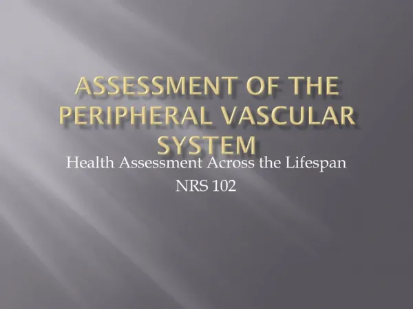
Assessment and Management of Patients With Vascular Disorders and Problems of Peripheral Circulation . Vascular System. Arteries and arterioles Capillaries Veins and venules Lymphatic vessels Function of the vascular system. Systemic and Pulmonary Circulation . Peripheral Blood Flow.

E N D
Assessment and Management of Patients With Vascular Disorders and Problems of Peripheral Circulation
Vascular System • Arteries and arterioles • Capillaries • Veins and venules • Lymphatic vessels • Function of the vascular system
Peripheral Blood Flow • Flow rate = ΔP/R • Movement of fluid across the capillary wall; hydrostatic and osmotic force • Hemodynamic resistance • Blood viscosity • Vessel diameter • Regulation of peripheral vascular resistance
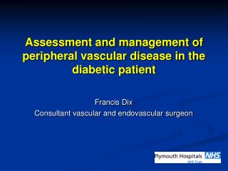
Assessment • Characteristics of arterial and venous insufficiency • Intermittent claudication • Rest pain • Changes in skin and appearance • Pulses • Aging changes
Continuous-wave Doppler ultrasound detects blood flow, combined with computation of ankle or arm pressures; this diagnostic technique helps characterize the nature of peripheral vascular disease.
ABI interpretation: ABI=1 normal (no arterial insufficiency) ABI= 0.95 mild arterial insufficiency ABI=0.5 moderate ABI< 0.5 ischemic rest pain ABI<0.25 sever ischemia (tissue loss)
Nursing Process: The Care of the Patient with Peripheral Arterial Insufficiency—Assessment • Health history • Medications • Risk factors • Signs and symptoms of arterial insufficiency • Claudication and rest pain • Color changes • Weak or absent pulses • Skin changes and skin breakdown
Nursing Process: The Care of the Patient with Peripheral Arterial Insufficiency—Diagnoses • Altered peripheral tissue perfusion • Chronic pain • Risk for impaired skin integrity • Knowledge deficient
Nursing Process: The Care of the Patient with Peripheral Arterial Insufficiency—Planning • Major goals include increased arterial blood supply, promotion of vasodilatation, prevention of vascular compression, relief of pain, attainment or maintenance of tissue integrity, and adherence to self-care program.
Improving Peripheral Arterial Circulation • Exercises and activities: walking, isometric exercises. Note: consult primary health care provider before prescribing an exercise routine • Positioning strategies • Temperature; effects of heat and cold • Stop smoking • Stress reduction
Maintaining Tissue Integrity • Protection of extremities and avoidance of trauma • Regular inspection of extremities with referral for treatment and follow-up for any evidence of infection or inflammation • Good nutrition, low-fat diet • Weight reduction as necessary
Nicotine Diet Hypertension Diabetes Obesity Stress Sedentary lifestyle C-reactive protein Hyperhomcysteinemia Age Gender Familial predisposition/genetics Risk Factors for Atherosclerosis and PVD Modifiable Nonmodifiable
Medical Management • Prevention • Exercise program • Medications • Pentoxifylline (Trental) and cilostazol (Pletal) • Use of antiplatelet agents • Surgical management
Medical management • Trental (pentoxifylline): increase erythrocyte flexibility, reduce blood viscosity, and has antiplatlet effect. • Pletal (cilostazol): decrease platelets aggregations, inhibit smooth muscles cell proliferations increase vasodilatations. • Anti-platelets aggregating agents (aspirin, clopidogrel (Plavix)): prevent the formation of thromboemboli
Surgical managements • Amputations (if occlusion is sever) • Vascular grafting (anastemosis) depends on the degree and location of stenosis or occlusion. • Endarterectomy: thrombus that obstruct the artery removed through incision to the artery affected.
Venous Thromboembolism • Pathophysiology • Risk factors • Endothelial damage • Venous stasis • Altered coagulation • Manifestations • Deep veins • Superficial veins
Pathophysiology • The exact cause is not known, but three reasons are known called Virchow’s triad: stasis of blood (venous stasis), vessel wall injury, and altered blood coagulation. • Thrombophelibitis: • Phlebothrombosis:stasis or hypercoagulability but without inflammation.
Blood flow and function of valves in veins. Note impaired blood return due to incompetent valve.
Clinical Manifestation • Deep veins: • Edema and swelling of extremities • Warm (affected extremity) • Superficial vein appears more prominent • Tenderness • +vehoman’s sign (not specific) • Superficial veins: • Pain or tenderness, redness, and warmth. • Can be treated with bed rest, leg elevations, analgesics, and anti-inflammatory drug.
Diagnosis: 1. Venography: The radiologist injects contrast material into a vein on the top of the foot. The blood clot appears as a defect in contrast material on the X-ray picture of the veins. 2. Duplex ultrasound: noninvasive procedure reflects gray-scale imaging for vein or artery. Help in determination the level and extent of venous disease and locate the disease stenosis or occlusion
Preventive Measures • Elastic hose • Pneumatic compression devices • Subcutaneous heparin, warfarin (Coumadin) for extended therapy • Positioning: periodic elevation of lower extremities • Exercises: active and passive limb exercises, and deep breathing exercises • Early ambulation • Avoid sitting/standing for prolonged periods; walk 10 minutes every 1-2 hours.
Nursing Process: The Care of the Patient with Leg Ulcers—Assessment • History of the condition • Treatment depends upon the type of ulcer • Assess for presence of infection • Assess nutrition
Arterial Ulcer, Gangrene Due to Arterial Insufficiency, and Ulcer Due to Venous Stasis
Medical Management • Anti-infective therapy is dependent upon infecting agent • Oral antibiotics are usually prescribed. • Compression therapy • Debridement of wound • Dressings • Other
Nursing Process: The Care of the Patient with Leg Ulcers- Diagnoses • Impaired skin integrity • Impaired physical mobility • Imbalanced nutrition
Collaborative Problems/Potential Complications • Infection • Gangrene
Nursing Process: The Care of the Patient with Leg Ulcers—Planning • Major goals include restoration of skin integrity, improved physical mobility, adequate nutrition, and absence of complications.
Mobility • With leg ulcers, activity is usually initially restricted to promote healing • Gradual progression of activity • Activity to promote blood flow; encourage patient to move about in bed and exercise upper extremities • Diversional activities • Pain medication prior to activities
Other Interventions • Skin integrity • Skin care/hygiene and wound care • Positioning of legs to promote circulation • Avoidance of trauma • Nutrition • Measures to ensure adequate nutrition • Adequate protein, vitamin C and A, iron, and zinc are especially important for wound healing • Include cultural considerations and patient teaching in the dietary plan
Varicose Veins (Varicosities) • Are abnormally dilated, tortuous, superficial veins caused by incompetent venous valves • Occurs in lower extremities, in the saphenous system or the lower trunk • Correlated with ↑ age, most in women, and people with occupation required prolonged standing • Other factors that cause VV are: hereditary, pregnancy
Pathophysiology: Primary: without involvement of deep veins) Secondary: resulting from obstruction of deep veins Reflux of venous blood result in venous stasis Clinical Manifestations: Dull aches muscle cramps ↑ muscle fatigue in lower legs Ankle edema Feeling of heaviness of the legs If deep veins obstructed pt will have S&S of chronic venous insufficiency (edema, pain, pigmentation, ulceration) Increased susceptibility to infection and injury.
Dx test is duplex scan ( document the anatomic site of reflux and provide a measure for the severity of valvular reflux
Prevention: Avoid activity that cause venous stasis as ( wearing constrictive clothing, crossing the legs, sitting or standing for long periods) Change position frequently Elevating the legs Walking 1-2 miles each day Elastic stoking Control wt.
Medical Management • Ligation and stripping: is done for primary VV, deep veins should be patent. Saphenous vein ligated in the groin where the saphenous vein meets the femoral vein, then 2-3 incision is made below the knee, stripper( wire) is inserted to the point of ligation, the wire is then withdrawn and vein as it is removed. • Thermal ablation • sclerotherapy
Nursing Management After surgery: • Bed rest is discouraged and early ambulation is encouraged • Instruct pt to walk Q one hour for 5-10min while awake for the 1st 24hr, then ↑ activity as tolerated • Wear elastic stocking continuously for 1wk • Elevate foot of bed • Standing and sitting are discouraged
Promote comfort and understanding: give analgesic, inspect dressing for bleeding, alert for reported sensations of “pins and needles.” Hypersensitivity to touch in the involved extremity may indicate a temporary or permanent nerve injury resulting from surgery The patient is instructed to dry the incisions well with a clean towel using a patting technique, rather than rubbing The patient is instructed to apply sunscreen or zinc oxide to the incisional area prior to sun exposure If the patient underwent sclerotherapy, a burning sensation in the injected leg may be experienced for 1 or 2 days
Cellulitus and Lymphatic Disorders • Cellulitus: infection and swelling of skin tissues • Lymphangitis: inflammation/infection of the lymphatic channels • Lymphadenitis: inflammation/infection of the lymph nodes • Lymphedema: tissue swelling related to obstruction of lymphatic flow • Primary: congenital • Secondary: acquired obstruction