Viral Hepatitis
Viral Hepatitis. Aswad H. Al.Obeidy FICMS, FICMS GE&Hep Kirkuk General Hospital. Hepatitis A. In 1982, HAV was classified as an enterovirus type 72 belonging to the Picornaviridae family


Viral Hepatitis
E N D
Presentation Transcript
Viral Hepatitis Aswad H. Al.Obeidy FICMS, FICMS GE&Hep Kirkuk General Hospital
Hepatitis A • In 1982, HAV was classified as an enterovirus type 72 belonging to the Picornaviridae family • Subsequent determination of the sequence of HAV nucleotides and amino acids led to questioning of this classification, and a new genus, Hepatovirus, was created for HAV • HAV has an icosahedral shape and lacks an envelope • It measures 27 to 28 nm in diameter • Inactivated at 85°C for 1 minute • Only one serotype of HAV is known • The HAV genome consists of a positive-sense RNA that is 7.48 kb long, single-stranded, and linear
EPIDEMIOLOGY • The epidemiologic risk factors for HAV infection reported for the U.S. population in 2002 were as follows: • Unknown, 57%; • Sexual or household contact with a patient who has hepatitis A, 12%; • International travel, 9%; • Male homosexual activity, 8%; • Injection drug use, 5%; • Child or employee in a daycare center, 1%; • Food or waterborne outbreak, 1%; • Contact with a daycare child or employee, 3%; • Other contact with a patient who has hepatitis, 4%
PATHOGENESIS • Once HAV is ingested and survives gastric acid, it traverses the small intestinal mucosa and reaches the liver via the portal vein • Once the virus reaches the hepatocyte, it starts replicating in the cytoplasm • Once the virus is mature, it reaches the systemic circulation via the hepatic sinusoids and is released into the biliary tree through bile canaliculi, passed into the small intestine, and eventually excreted in the feces • The pathogenesis of HAV-associated hepatocyte injury is not completely defined • Immunologically mediated cell damage is more likely
CLINICAL FEATURES • HAV infection usually presents in one of the following five different clinical patterns: • (1) asymptomatic without jaundice • (2) symptomatic with jaundice and self-limited to approximately 8 weeks • (3) cholestatic, with jaundice lasting 10 weeks or more • (4) relapsing, with two or more bouts of acute HAV infection occurring over a 6- to 10-week period • (5) FHF
EXTRAHEPATIC MANIFESTATIONS • Consist most commonly of an evanescent rash (14%) • Arthralgias (11%) • Less commonly of leukocytoclastic vasculitis • Glomerulonephritis • Arthritis • In rare cases, hepatitis A has been followed by the development of type 1 AIH
DIAGNOSIS • A diagnosis of acute hepatitis A requires demonstration of IgM anti-HAV in serum • The test result is positive from the onset of symptoms and usually remains positive for approximately 4 months • Some patients may have low levels of detectable IgM anti-HAV for more than a year after the initial infection • IgG anti-HAV is also detectable at the onset of the disease, remains present usually for life, and, after clinical recovery • IgG anti-HAV interpreted as a marker of previous HAV infection
TREATMENT • There are no specific medications to treat acute hepatitis A • Symptomatic treatment is the rule • Attention to sanitation and administration of serum immune globulin (IG) are the mainstays of preventing HAV infection • The availability of excellent HAV vaccines has rendered use of IG for preexposure prophylaxis unnecessary • When IG is used for postexposure prophylaxis, it should be given within 2 weeks of exposure • In these cases, the recommended dose is 0.02 mL/kg, given by intramuscular injection • Although considered safe, IG can cause fever, myalgias, and pain at the injection site • Postexposure prophylaxis with IG can be accompanied safely at the same time as initiation of active immunization with the vaccine
HEPATITIS B • An estimated 400 million persons are carriers of hepatitis B virus (HBV) in the world today • Effective vaccines against HBV have been available for more than 20 years • Hepatitis B is the chief cause of cirrhosis and HCC in the world today • The virus is 100 times as infectious as human immuno-deficiency virus (HIV) and 10 times as infectious as hepatitis C virus (HCV) • One or more serologic markers of HBV infection were demonstrated in 4.9% of the U.S. population, and the prevalence of chronic infection was 0.2%
Definitions • HBV carrier has often been used to refer to persons persistently infected with HBV who have normal serum aminotransferase levels • Inactive carriers are patients who have evidence of HBV replication on a PCR-based assay only • Long-term follow-up of inactive carriers suggests that the majority of these patients do not have progressive liver disease and do not experience complications • Active carriers, on the other hand, have evidence of HBV replication on non–PCR-based assays for HBV DNA, intermittently or persistently elevated serum aminotransferase levels, and evidence of chronic hepatitis in a liver biopsy specimen
Clinical Sequelae of Acute Hepatitis B Virus Infection • The age at which a person becomes infected with HBV is a principal determinant of the clinical outcome • HBV infection in adults with intact immune systems is likely to cause clinically apparent acute hepatitis B; only 1% to 5% of these persons become chronically infected • By contrast, as many as 95% of infected neonates become chronic HBV carriers because of immunologic tolerance to the virus. • In adults, fulminant liver failure due to acute hepatitis B occurs in less than 1% of cases • The rate of spontaneous survival in acute liver failure due to hepatitis B is only approximately 20%
CLINICAL FEATURES • The incubation period of acute hepatitis B varies from a few weeks to 6 months (average, 60 to 90 days) • Acute infections are heralded by a serum sickness–like prodrome of fever, arthralgias/arthritis, and rash, which is most commonly maculopapular or urticarial, in 10% to 20% of patients • These features generally abate before the manifestations of liver disease and peak serum aminotransferase elevations are observed • Jaundice develops in only about 30% of patients • Clinical symptoms and jaundice generally disappear after 1 to 3 months • Persistence of HBsAg after six months implies development of a carrier state • Serum aminotransferase levels of 1000 to 2000 U/L are typical
Extrahepatic Manifestations • Arthritis • Dermatitis • Glomerulonephritis • Polyarteritis nodosa • Mixed cryoglobulinemia • Papular acrodermatitis • Polymyalgia rheumatica
DIAGNOSIS • HBsAg appears in serum 2 to 10 weeks after exposure to HBV • The disappearance of HBsAg is followed several weeks later by the appearance of anti-HBs. In most patients, anti-HBs persists for life and provides long-term immunity • Anti-HBc is detectable in acute and chronic HBV infection. During acute infection, anti-HBc is predominantly of the IgM class and is usually detectable for 4 to 6 months after an acute episode of hepatitis • The finding of HBeAg in the serum of an HBsAg carrier indicates greater infectivity, a high level of viral replication, and the need for antiviral therapy • The measurement of serum HBV DNA is commonly employed to evaluate a patient's candidacy for antiviral therapy and to monitor response during treatment
TREATMENT • All the guidelines indicate that treatment decisions ideally should be made in the context of liver histologic findings and that treatment should be directed preferentially to patients with histologically moderate to severe hepatitis • Currently, treatment decisions are not based on specific grading or staging of liver histology • Interferon Advantages Finite duration of treatment. Durable off-treatment response. Loss of HBsAg (5%-8%) Disadvantages Given by injection. Frequent side effects. Expensive. Low response rate with a high level of viremia • Nucleoside analogs Advantages Negligible side effects. Potent inhibition of virus replication. Less expensive than interferon. Disadvantages Oral delivery. Drug resistance. Long or indefinite treatment duration. Low rate of HBsAg disappearance. Moderately expensive when given long term
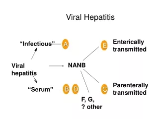
HEPATITIS D (DELTA) • Hepatitis D (delta) virus was discovered by Rizzetto and associates in 1977 as a unique nuclear antigen in the hepatocytes of patients infected with HBV • At least 5% of HBV carriers worldwide are estimated to be infected with HDV, and therefore, the overall burden of HDV infection is between 15 and 20 million cases • Among the three genotypes of HDV (I to III), genotype I is the most prevalent and is the most common genotype in Mediterranean countries, Africa, Europe, and North America • Genotype II is reported mostly in Japan and Taiwan and is associated with milder liver disease • Genotype III has been isolated from epidemics in South America
PATHOGENESIS • The pathogenesis of HD-related liver disease depending on the interplay of three major factors: (1) HDV-associated factors, such as genotype 2 and the expression of specific HDAg species SHDAg, but not LHDAg, is directly cytotoxic to hepatocytes. (2) Host associated factors, such as the immune response. Several autoantibodies have been described in association with chronic HDV infection. The major LKM-3 antigen appears to be a family of uridine diphosphate-glucuronosyltransferases (UGTs), which are hepatic enzymes involved in phase 2-drug metabolism. (3) Helper virus-associated factors, such as the HBV genotype and the level of HBV replication
CLINICAL FEATURES • The clinical and laboratory findings vary with the type of infection: • (1) Coinfection of HBV and HDV results in acute hepatitis B+D with high incidence of liver failure, the rate of progression to chronic infection is not different from that after classical acute hepatitis B • (2) Superinfection with progression to chronic HDV infection occurs in almost all patients • (3) Helper independent latent infection, which can be rescued by the helper virus at a later time
TREATMENT • The management of HDV infection is supportive • Liver transplant is the treatment of choice for fulminant or end stage liver disease • The only drug that has been examined in randomized controlled trials is interferon • HBV/HDV coinfection can be prevented by either preexposure or postexposure prophylaxis for HBV • While prevention of HDV superinfection depends primarily on behavior modification
Hepatitis C • HCV is a single-stranded positive-sense RNA virus that belongs to the Flaviviridae family • Hepatitis C virus (HCV) infection is the most prevalent bloodborne infection in the United States, affecting an estimated 4 million persons • Most patients acutely infected with HCV are asymptomatic and do not clear the infection spontaneously; approximately 85% develop chronic infection • Of patients with chronic infection, 20% to 25% develop cirrhosis over 20 years • Of patients who develop cirrhosis, the risk for hepatocellular carcinoma is approximately 5% per year
DIAGNOSIS • The anti-HCV antibody test is the screening test for at-risk persons • A positive test in a person with one of the risk factors confirms exposure to the virus • The HCV RNA test is required to determine active infection rather than just exposure to the virus • In patients with none of the risk factors who have a positive anti-HCV antibody test and a negative HCV RNA test, the recombinant immunoblot assay (RIBA), which detects antibodies to any of several viral antigens, should be done • A positive RIBA, in the absence of detectable HCV RNA confirms that the patient has been infected with hepatitis C but has spontaneously cleared the infection
TREATMENT • Therapy for hepatitis C infection consists of the combination of pegylated interferon and ribavirin • The ideal candidate for therapy is the patient with detectable virus, some indication of hepatic inflammation (elevated liver tests or inflammation on the biopsy), and no contraindication to therapy • The goal of therapy is to achieve a sustained virologic response, which is defined as undetectable HCV beyond 6 months after the end of treatment • Eleven distinct genotypes of the virus have been identified • The most important variable affecting the response rate is the genotype of the virus • Patients who achieve at least a two-log reduction in the HCV RNA by the twelfth week have a higher likelihood (approximately 66%) of achieving a sustained response
Hepatitis E • Hepatitis E is a form of acute, icteric, self-limited viral hepatitis caused by the hepatitis E virus • The disease was first recognized in the 1980s • Its genome was cloned in 1990 and fully sequenced shortly thereafter • It results from fecal-oral transmission • Pregnant woman with acute HEV infection are at greatest risk for developing severe hepatitis or liver failure