Picornaviruses
Picornaviruses. Plus Strand RNA Virus Families. PICORNAVIRIDAE • More than 200 viruses prevalent world-wide. • cause many serious diseases of animals and man. • Foot and mouth virus first animal virus described (1898). • Poliovirus is an important model:

Picornaviruses
E N D
Presentation Transcript
Plus Strand RNA Virus Families PICORNAVIRIDAE • More than 200 viruses prevalent world-wide. • cause many serious diseases of animals and man. • Foot and mouth virus first animal virus described (1898). • Poliovirus is an important model: - first virus purified and crystallized. - first inactivated vaccine used (Salk 1950’s). - first picornavirus to be sequenced. - first infectious cDNA clone of an animal virus. - first picornavirus structure to be solved.
PICORNAVIRUS PROPERTIES • Capsids are small unenveloped 25 -30 nm icosahedra. • Resistant to pH 3 to 9 (except for Rhinoviruses). • Plus sense single stranded RNA genomes (~7400 bases). • Genome is monopartite. • RNA 5’ end has a covalently attached VPg (22-24 aa) and 3’ end is polyadenylated. • The 5’ end contains a highly structured (~740 nt) untranslated region that contains several AUG’s. • The naked RNA is sufficient for infection. • The RNA is translated into a polyprotein that is cleaved into enzymatic and structural proteins. • The Virus replicates in the cytoplasm.
Members of the Picornaviridae Cause Many Serious Diseases of Man and Animals Genus Enterovirus - Poliovirus type member, 3 major types cause paralysis. Genus Rhinovirus - cause respiratory tract infections, acid labile, cause colds in humans (110 types) and pigs. Genus Hepatovirus - Hepatitis A, contagious liver infections. Genus Cardiovirus - EMC group, cause heart and brain inflammation acid labile, source is a rodent reservoir. Genus Apthovirus - Foot and mouth disease, most destructive in Africa.
5-fold Axis 3-fold Axis 2-fold Axis VP1 VP2 VP3 PICORNAVIRUS STRUCTURE • The Basic capsid building block is a protomer that consists of one copy each of VP1, VP2, VP3 & VP4. • VP1, VP2 & VP3 are on the virion surface, with VP4 being internal. • VP1, VP2 & VP3 have no sequence homology, but have the same topology. • Five protomers assemble into a pentamer. • The icosahedron is formed by 12 pentamers.
Picornavirus Capsid Structure Capsid is a pseudo T=3 icosahedron consisting of 60 identical asymmetric protomers arranged as 12 pentamers. Each protomer is composed of a single copy of each of the four capsid proteins, VP1, VP2, VP3 and VP4. VP4 is located on the inner surface of the protein shell formed by VP1, VP2 and VP3. All three proteins contain a central b–barrel jelly roll. The jelly roll is a wedge-shaped structure that consists of two antiparallel b-sheets.
Picornavirus Members Bind to Many Different Host Receptors IG Like - Members of the immunoglobulin family of receptors found on cells of the immune system. Scr-short consensus repeat LDL-low density lipoprotein T/S/P threonine/serine/proline
A single type of receptor mediates virus Human Rhinovirus binding and entry • The integral membrane protein, intercellular adhesion molecule 1 (Icam-1) was identified as the receptor for the major group of human rhinoviruses. • Icam-1 is a member of the immunoglobulin superfamily. • Icam -1 is found on the surface of many tissues, including nasal epithelium and lung epithelium. • The normal function of Icam-1 is to bind a ligand on the surface of lymphocytes and to promote immunological and inflammatory functions. • This host response accounts in part for cold symptoms.
Picornavirus Attachment To the Icam-1 Receptor • A depression or canyon is formed at the junction of VP1 and VP3 in the capsids of rhinoviruses and some enteroviruses like poliovirus. • The canyons are the sites of interaction with cell surface receptors. • As the picornavirus binds to Icam-1, the host receptor penetrates into the viral canyon and this causes a change in conformation of the capsid to permit virus entry. • Antibody molecules can also penetrate into the canyon and antibodies that bind to the virus in this manner neutralize infectivity by blocking entry of the receptor into the canyon. However, viruses quickly mutate to change the shape of the canyon to prevent antibody binding. • Some antiviral drugs (WIN compounds) can irreversibly replace the lipid that lines the tunnel and inhibit uncoating and in some cases attachment of the virus.
Entry of Poliovirus into Cells • Nonenveloped poliovirus enters cells by forming a pore in the membrane of the cell. • During interactions of poliovirus with its receptor major conformational rearrangements occur in the virus particle. • The particles lose VP4 and the hydrophobic N-terminus of VP1 is displaced to the virion surface • N-termini of VP1 forms a pore in the cell mebrane through which the RNA is released into the cytosol. • Some evidence suggests that virus particles may undergo endocytosis in some cell types.
Poliovirus Genome organization •Poliovirus genome contains a single ORF, which encodes a 247 kD polyprotein. It has VPg at its 5’ end and a poly(A) tail at its 3’ end •Processing occurs in 3 steps. The first is to cleave the P1 capsid protein precursor, which is catalyzed by 2Apro • The second step is to process the noncapsid and the capsid precursors catalyzed by 3Cpro and 3CDpro • The third step is the processing of VP0 into VP4 and VP2
During translation, the 2A proteinase cleaves at its amino terminus immediately after it is translated. • The 2A cleavage releases the structural protein (P1) that is the precursor to the individual viral capsid proteins. • The Proteinase 3C and its precursor 3CD carry out the remaining polyprotein cleavages. • The 2B protein is a host range determinant that is also involved in RNA synthesis. • The 2C protein is very highly conserved and is also involved in RNA synthesis. • 3D is an RNA dependent RNA polymerase and has GDD polymerase motif. • The 3B protein is the VPg. Polyproteins are Proteolytic Processed During Translation
More Ribosomal Scanning!!!!!!! • eIF4G, a component of the eIF4F cap binding complex, acts as a bridge between the cap structure and the 40S subunit. • The helicase, eIF4A, associates with eIF4F to unwind secondary structure in the UTR. • As 40S subunit scans the untranslated Region (UTR) a number of eIFs (eukaryotic elongation factors) including eIF-3, eIF-2, eIF-4 & eIF-5 become associated with the subunit. • The 40S subunit plus the eIFs increase subunit size progressively. • eIF-5 associates with the subunits to promote 60S subunit assembly and recognition of AUG Kozak Sequences(5’-GCA/GCCAUGG-3’).
Picornavirus Translation: • Picornavirus translation is cap independent • Picornavirus mRNAs have no cap (pUp) at their 5’ terminus and no VPg. • The genome has a (743 nt) long UTR untranslated leader sequence that contains 8 upstream AUGs preceding the translational initiation site. • Internal ribosomal binding occurs at an Internal Ribosomal Entry Sequence (IRES). • The IRES consists of a high level of secondary structure in the UTR leader sequence that mediates ribosome 40 S subunit binding and initiation. • eIF-3, eIF-4G and eIF-4a promote IRES assembly. A host factor X is also required.
Short 35S Methionine Pulse Protein Synthesis Is Bimodal in Infected Cells • Protein synthesis shows rapid decline 30 min postinfection. • After two hours synthesis is lowest but increases rapidly. • By four hours most synthesis is poliovirus protein. • Synthesis again declines rapidly near end of replication.
Mechanisms for Inactivating Eif-44 Activity Poliovirus 2A protein Dephosphorylates eIF4Ebp and Cleaves the eIF4G subunit.
Replication of Picornavirus RNA P gRNA - RNA mRNA • The first step in replication is to translate the viral RNA. • Picornaviruses replicate using an RNA copying mechanism. • The replicating RNA has a VPg, but the mRNAs lack this protein.
Poliovirus Replication initiates by formation of a Covalent Bond • Polioviral RNA is linked to the 22 aa VPg via a uridine- tyrosine phosphodiester bond. • During replication, mRNAs are produced by cleavage of this phosphodiester bond by a cellular enzyme to produce viral mRNAs containing a 5’ terminal Up. • Genomic RNAs incorporated into virus particles contain the 5’ VPg. • These steps occur in vesicles derived from the smooth endoplasmic reticulum.
A Host Protein is Required for Synthesis of Polio Virus RNA A highly based paired structure formed from the 108 nucleotides at the 5’ end of the (+) strand RNA of poliovirus, which forms a cloverleaf like structure. The viral protein 3CD and a PCBP a host Cellular protein, (PolyrC Binding Protein) interact with different loops of the cloverleaf. This interaction brings the 5’ end close to the 3’ end. At this point VPg priming can occur. Upon completion of the minus strand a similar clover leaf is formed at the 5’ end of the minus strand. Formation of this ribonucleoprotein complex is required for synthesis of both plus and minus strand RNA.
Minus strand synthesis: • Precursor of VPg, 3AB, acts as a VPg donor • A ribonucleoprotein complex is formed when PCbp and 3CDpro bind to the cloverleaf structure • This complex interacts with PAbp1, producing circular genome
Protease 3CDpro cleaves membrane bound 3AB to produce VPg • VPg is uridylated by 3Dpol using the cre sequence as a template and transferred to the 3’ end of the genome • 3D pol uses uridylated VPg as a primer for (–) strand RNA synthesis
Plus strand synthesis: • The strands of the RF • are separated by 2C, • which binds to cloverleaf • in the (-) strand • Membrane bound 3AB is • cleaved to produce VPg • Uridylated VPg is • synthesized by 3D pol, • using 3’ terminal A residues • of the (–) strand RNA as a • template • Uridylated VPg is then • elongated by 3D pol to • synthesize (+) strand RNA
P1 Peptide Folded P1 5S Unit Steps in the Assembly of Poliovirus • After Translation and release by the 2A protease, P1 folds and is cleaved by the 3CD protease to form the 5S protomers. • Pentamers of the 5S units assemble into a 14S complex. • The 14S complex may then form a 75S empty capsid that assembles with the RNA to form a noninfectious 150S provirion. • Some researchers argue that the 14S subunits assemble in a series of steps with the viral RNA to form provirions. • The provirion is converted to an infectous form by internal cleavage of VP0 molecules to VP4 plus VP2. Assembly steps are not reversible
Poliovirus Life Cycle The capsid binds to host receptors and releases Viral RNA into the cell. Translation of the released RNA produces a polyprotein that is processed by the 2A and 3CD proteinases to form the viral gene products. RNA synthesis is initiated by the 3AB proteins and elongated by the 3D protein. As proteins viral capsid assembly begins in the cytoplasm from P1 products.
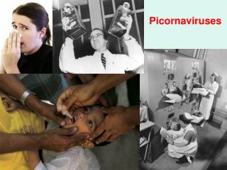
Disappearance of Polio in the USA In 1952 there were 57,000 cases of polio in USA mostly in children between five to nine years old. By 1961 less than 100 cases were reported & by 1970 less than 10 cases occurred per year.
Science 296:356 (April 12, 2002) Live Vaccine induced cases of polio have occurred. Now the eradication programs are switching back to the killed virus vaccine.
Flaviviruses • • Flavivirus virions have enveloped particles. The enveloped icosahedral virions are composed of a lipid bilayer surrounding an icosahedral nucleocapsid. The envelope protein forms a very tight protective shell composed of 90 E dimers (180 subunits) that bind close to the inner capsid protein. • • Genome is a single stranded positive sense RNA with a cap structure at the 5’ end. The 3’ end of the flaviviruses lack a poly(A) tail. Thus, the flaviviruses differ from picornaviruses at their 5’ and 3’ terminus. • • The viral proteins are produced from a single polyprotein that is cleaved by host and viral proteases. Thus the flavivirus protein expression strategy has some similarities to the expression strategies of the Picornaviridae • Transmitted mostly by mosquitos or ticks
Flavivirus Genome Organization and Protein Functions Structural Polymerase Genes C - Nucleocapsid protein that forms the enveloped icosahedron. prM - Structural glycoprotein. Cleaved to pr and Membrane protein by furin. E - Envelope Protein NS1 - Nonstructural glycoprotein required for RNA replication. NS2A - Hydrophobic protein that anchors replication machinery in the Endoplasmic Reticulum. NS2B - Hydrophobic protein. Serves as cofactor for the NS3 protease. NS3 - Serine protease; Helicase; component of capping enzyme. NS4A & B - Hydrophobic proteins that may anchor replication machinery in the Endoplasmic Reticulum. NS5 - RNA polymerase contains capping enzyme activity.
Flavivirus Envelope Protein Translation and Processing • A transmembrane insertion occurs at the ER during viral protein translation. • The capsid protein has a hydrophobic signal sequence at its C-terminus that translocates the prM (membrane precursor protein) into the ER lumen. • The viral NS3 protease cleaves the Capsid from the prM. • The capsid protein associates with viral RNA and assembles at the ER surface as an immature virion.
The immature virion moves to the Golgi where the prM and envelope proteins are glycosylated and the envelope protein and a nonstructural protein (NSI) are trimmed by Host Signalase enzymes. • A host protein (Furin) cleaves the prM protein late in viral maturation.