Download

1 / 25
420 likes | 2.35k Vues
Tests for the Evaluation of Lupus Anticoagulants . Islamic University of Gaza. Lupus Anticoagulants . General Background

E N D
Tests for the Evaluation of Lupus Anticoagulants Islamic University of Gaza
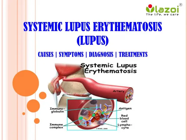
Lupus Anticoagulants General Background • Lupus anticoagulants (LA) were first described in the year 1952 by Conley and Hartmann in two patients suffering from Systemic Lupus Erythematosus (SLE). It was given the name “Lupus” since it was initially recognized in patients with SLE and the name “Anticoagulant” because the two patients diagnosed as SLE developed bleeding complications. • It is now clear that Lupus anticoagulant is also encountered in patients without SLE and is associated with thrombosis rather than abnormal bleeding. • Thus LA is neither a syndrome nor a disease but a laboratory phenomenon detected by prolonged results with phospholipid dependent clot based assays.
Lupus Anticoagulants • Lupus anticoagulants are antibodies directed against several types of phospholipid-protein complexes. • Lupus anticoagulants impair the in vitro phospholipid dependent activation of factor X, factor IX, and prothrombin. • Consequently, the APTT is prolonged in the presence of a lupus anticoagulant. For this reason, most laboratories use the APTT as their primary lupus anticoagulant screen. Because they have a variety of targets.
Lupus Anticoagulants • lupus anticoagulants are called nonspecific inhibitors. • Chronic lupus anticoagulants confer a 30% risk of arterial or venous thrombosis; thus every acute care laboratory must provide a means for their detection. • The presence of an LA is usually not associated with a bleeding problem unless accompanied by • thrombocytopenia, • factor II deficiency, • platelet dysfunction • Or drug administration (e.g., aspirin).
Scientific Sub - comittee criteria for the laboratory diagnosis of Lupus anticoagulants* IN order to make a diagnosis of LA; sample should show each of the following: • Prolongation of at least one phospholipid – dependent clotting assay. • Evidence of inhibitory activity shown by the effect of patient plasma on pooled normal plasma. • Evidence that the inhibitory activity is dependent on concentration of phospholipid. This may be achieved by addition or alteration of phospholipids/ hexagonal phase phospholipids/ platelet/ platelet vesicles in the test system. • LAs must be carefully distinguished from other coagulopathies that may give similar laboratory results or may occur concurrently with LAs. Specific factor assays and the clinical history may be helpful in differentiating LAs from these other possibilities.
Laboratory Diagnosis of Lupus Anticoagulants • The traditional screening methods used for the laboratory diagnosis of Lupus • Anticoagulants can be broadly classified as follows: • Immunological assays • Clot based assays incorporating phospholipids in the reagent sysytem
A. Immunological Assays Anti-Phospholipid Antibody Assays Principle • Anti-beta-2 glycoprotein I (anti-β2GPI) and Anticardiolipin antibodies (ACAs) are antiphospholipidimmunoglobulins that are IgG, IgM, IgA, or a combination. • Antiphospholipidantibodies are a heterogeneous group of autoantibodies including ACA, LA, beta-2 glycoprotein-l (β2GPI), antiprothrombin, and antiphosphatidylserine(APTS). • Some patients with elevated anti-β2GPI and/or ACAs have been reported to also have an LA.
Anti-Phospholipid Antibody Assays • Several studies have shown that patients with the LA and the closely related anti-β2GPI/ACA are prone to • recurrent venous and arterial thrombosis, • recurrent spontaneous abortions, • thrombocytopenia. This tendency has been described as the antiphospholipid syndrome (APS). • The criteria for diagnosing this syndrome are divided into clinical and laboratory groups.
Anti-Phospholipid Antibody Assays • APSis present if at least one of the clinical criteria and one of the laboratory criteria that follow are met: • Clinical criteria: vascular thrombosis or pregnancy morbidity • Laboratory criteria: • Lupus anticoagulant present in plasma on two or more occasions at least 12 weeks apart. • Anticardiolipinantibody, IgG or IgM positive in medium or high titer on two or more occasions at least 12 weeks apart • Anti-β2GPl antibody, IgG or IgM present on two or more occasions at least 12 weeks apart. • The positive laboratory criteria and the clinical criteria should occur within 5 years of each other.
A. Immunological Assays • Individuals with Lupus anticoagulants may also show the presence of other anti phospholipid antibodies. The most frequent finding is the presence of Anti- cardiolipin antibodies. • The commonly employed method is the ELISA technique where the solid phase is coated with cardiolipin and β-2 GPI (as a cofactor). The ELISA method detects IgM, IgG and IgA class of anti-cardiolipin antibodies. • An important point to note is that Lupus anticoagulants and anti-cardiolipin antibodies differ in their phospholipid-binding characteristic hence detection of anti cardiolipin antibodies is not specific for the presence of Lupus anticoagulants though they may be present in the same patient.
B. Clot based assays • APTT ( Activated Partial Thromboplastin Time) • TTI (Tissue Thromboplastin Inhibition test) • KCT (Kaolin Clotting Time) • PNP (Platelet Neutralization procedure) • Hexagonal Phase Phospholipids (Staclot-LA)
APTT ( Activated Partial Thromboplastin Time) Since Lupus anticoagulants bind to the phospholipid complex they do prolong phospholipid based coagulation assays. Logically the activated partial thromboplastintime (APTT) is prolonged and this property has been used for the detection of LA using APTT reagents. In the context of LA detection, the APTT test has certain shortcomings: • An important variable related to the suitability of APTT reagents in the detection of Lupus anticoagulants is the composition of phospholipids used in the reagent system. Different reagents have varying sensitivity for the presence of Lupus anticoagulants on the basis of their phospholipid composition.
APTT ( Activated Partial Thromboplastin Time) • The APTT test is affected by the presence of inhibitors to Factor VIII, IX and XI. In order to rule out the presence of inhibitors usually mixing studies are performed. In mixing studies a 1:1 or 1:4 ( Normal plasma: Patient plasma) is used. Failure to correct the prolongation of clotting time using the mixing studies indicates the presence of Lupus anticoagulants. • The APTT test is also the test of choice for monitoring heparin therapy. This reduces the specificity of the APTT to LA. The presence of heparin can be ruled out by using a thrombin time test. If thrombin time test shows normal values, then the sample does not contain heparin. But if the thrombin time test is abnormal, then heparin neutralization test is used which includes protamine sulphate. In this test, various concentrations of protamine sulphate are added to plasma before the addition of thrombin reagent. When protamine sulphate neutralizes all the heparin present, the clotting time reverts to normal value.
TTI (Tissue Thromboplastin Inhibition test) • The Tissue Thromboplastin Inhibition test utilizes a diluted PT reagent. The results are expressed as ratio of patient values to normal control values. • The TTI test is affected by numerous variables: • Species and tissue origin of thromboplastin can affect the test results as different sources of thromboplastin have varying sensitivity and responsiveness. • Choice of “Normal” reference plasma is the most critical variable, because depending on the laboratory the choice of reference plasma could be lyophilized plasma, a frozen plasma pool or fresh plasma. The ratio of patient to normal can therefore change according to the choice of “normal” plasma. • Some IgM Lupus anticoagulants do not prolong the TTI test
KCT (Kaolin Clotting Time) KCT is similar to APTT, the difference being that KCT reagent is devoid of phospholipids and incorporates Kaolin as contact activator. The test is performed on a range of mixture of normal and patient’s plasma. Different patterns of response are obtained indicating the presence of Lupus anticoagulants or the deficiency of one or more coagulation factors. The KCT test though sensitive is not specific for LA, additionally: • It cannot be automized • The test shows prolonged results with factor VIII, IX, XI & XII deficiency or corresponding inhibitors • The test is also highly sensitive for the presence of heparin.
4. PNP (Platelet Neutralization procedure) Principle • The platelet neutralization procedure (PNP) is based on the ability of platelets to significantly correct in vitro coagulation abnormalities. • The disrupted platelet membranes present in the freeze-thawed platelet suspension neutralize phospholipid antibodies present in the plasma of patients with LA. • After the patient plasma is mixed with the freeze-thawed platelet suspension, the APTT will be shortened when compared with the original baseline APTT. But if an inhibitor is directed against specific coagulation factor, the clotting time is not shortened.
4. PNP (Platelet Neutralization procedure) Interpretation • A correction of the baseline APTT of a defined amount of time (i.e., 3 to 5 seconds or more) by the platelet suspension as compared with the control is indicative of the presence of an LA. NOTE The PNP test though useful did not gain wide usage : • Due to limited stability the platelet preparations loose their activities on storage hence do not show reproducible results. • They cannot differentiate between Lupus anticoagulants and Factor VIII inhibitors.
Hexagonal Phospholipid Neutralization Assay Principle • The hexagonal phospholipid neutralization assay uses the same principle as the PNP assay, normalization of the aPTT in the presence of added phospholipid, but this assay specifically uses a phospholipid in a hexagonal conformation, Neutralization by this hexagonal form in an assay with a very lupus-anticoagulant sensitive aPTT reagent, is a more sensitive confirmation test than the PNP. Comment • Specimen collection, centrifugation, and processing are critical when testing for the presence of an LA.
Confirmatory Tests for Lupus Anticoagulants • Confirmatory tests to identify an LA include those that utilize a low concentration of phospholipid in the test system, thereby increasing the LA effect such as • the tissue thromboplastin inhibition test (TTIT), • dilute Russell's viper venom time (dRVVT), • and the kaolin clotting time (KCT), or • those that increase the phospholipid, thereby neutralizing the LA effect, such as the platelet neutralization
DiluteRussell’sViperVenomTime(DRVVT) • dRVVT : The test of choice for screening and confirmation of LA • Indicating the phospholipid dependence of LA • Achieving maximum sensitivty for the precence of LA’s. • In general dRVVT based tests comprise of: • SCREENING REAGENT, containing limited amount of phospholipids with RVV (Russell’s Viper Venom) • CONFIRMATION REAGENT, containing additional phospholipids with same amount of Russell’s Viper Venom, to confirm the presence of phospholipid dependent Lupus anticoagulants.
Principle of dRVVT for LA detection • Russell’s Viper Venom directly activates Factor V and X in presence of phospholipid and calcium ions, bypassing Factor VII of the extrinsic pathway and the contact and antihaemophilic factors of the intrinsic pathway. • In normal plasma, in the absence of lupus anticoagulants, Factor V and X is directly activated by Russell’s Viper Venom, which in presence of phospholipid and calcium ion leads to clot formation.
Principle of dRVVT for LA detection • In patients with LA, autoantibodies bind the epitopes of reagent phospholipids thereby preventing the activation of prothrombinase complex. This results in a prolongation of clotting time with SCREENING reagent. • The CONFIRMATION reagent incorporates additional phospholipids to neutralize the LA. Once LA are neutralized clot formation proceeds relatively uninterrupted achieving a lower clotting time, to prove the phospholipid dependence of the autoantibodies.
Interpretation of results with dRVVT test • If SCREEN TIME is prolonged, to confirm the presence of lupus anticoagulants the plasma sample is tested with CONFIRMATION REAGENT. • If CONFIRM TIME results shows a lower clotting time as compared to SCREEN TIME, it indicates the presence of phospholipid dependant Lupus Anticoagulants. • Also the results can be expressed as ratio, • The results expressed, as ratio is further useful in classifying the patient as normal, moderate, high and very high LA. • If results of the ratio are borderline, mixing studies may be done further with the sample specimen. The mixing studies should be carried out with a 50:50 mixture of test plasma and normal plasma.