Download
1 / 25
250 likes | 605 Vues
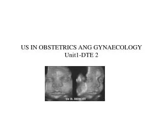
US IN OBSTETRICS ANG GYNAECOLOGY Unit1-DTE 2. CEPHALOMETRY. Inthis session Ultrasonic cephalometry To assess fetal gestational age. To monitor fetal growth by serial measurements. Technique BPD measurement Fetal body measurements Fetal abnormalities Anencephaly Hydrocephaly

E N D
CEPHALOMETRY Inthis session Ultrasonic cephalometry • To assess fetal gestational age. • To monitor fetal growth by serial measurements. Technique • BPD measurement Fetal body measurements Fetal abnormalities • Anencephaly • Hydrocephaly • Spina bifida • Hydramnios. • Fetal hydronephrosis
ANENCEPHALY • AnencephalyThis is where the brain does not develop properly or is absent, and the baby is either still born or dies shortly after birth. • Anencephaly is one of the most common neural tube defects. Neural tube defects are birth defects that affect the tissue that grows into the spinal cord and brain. • Anencephaly occurs early in the development of an unborn baby. • It results when the upper part of the neural tube fails to close. • occurs in about 1 out of 10,000 births. A front view of an anencephalic fetus
HYDROCEPHALY • HYDROCEPHALUS (from the Greek hydro = water, cephale = head). An accumulation of cerebrospinal fluid which arises from an imbalance in the production and drainage of that fluid. • Pathological basis Hydrocephalus is sometimes referred to as 'water on the brain'. A watery fluid, known as cerebro-spinal fluid or CSF, is produced continuously inside each of the four spaces or ventricles inside the brain. The CSF normally flows through narrow pathways from one ventricle to the next, then out across the outside of the brain and down the spinal cord. The CSF is absorbed into the bloodstream and recirculates. The amount and pressure are normally kept within a fairly narrow range. If the drainage pathways are blocked at any point, the fluid accumulates in the ventricles inside the brain, causing them to swell - resulting in compression of surrounding tissue. In babies and infants, the head will enlarge. In older children and adults, the head size cannot increase as the bones which form the skull are completely joined together.Most babies born with Spina Bifida also have Hydrocephalus
HYDROCEPHALY 3D Surface ReconstructionHydrocephalus Hydrocephaly at 18 week’s gestation age.displacement of the lateral wall of the lateral ventricles.
SPINA BIFIDA • Spina Bifida is one of the most common congenital defects, occuring within the first 25 days of pregnancy deformity also known as a neural tube defect. • An obvious gap in the skin covering the spine, the vertebrae and the nervous system is damaged,the child will have some degree of paralysis. • US interpretation- • Normal • fetal spine appears as a small closed ring to one side of fetal trunk. • Spina bifida • Spine appear kinked or incomplete in longitudnal section. • Spinal ring is incomplete posteriorly.
HYDRAMNIOS • Condition of excess liquor amnii • Often associated with anencephaly,multiple pregnancy or spina bifida.
CRL MEASUREMENT • Embryonic Crown–rump length(CRL) measurement-most accurate method of gestation age assessment in early pregnancy, made between 7 to 13 weeks • Definition: The measurement of the length of the unborn human fetus from the top of the head (crown) to the bottom of the buttocks (rump). • Serial CRL measurements give important information on early fetal development. • Following formula is an approximation: Gestational age (weeks) = crown-rump length (cm) + 6.5
CRL MEASUREMENT Gestational Sac size = 3 to 6mm At 4 weeks and 3 days, a tiny gestational sac becomes visible and grows at a rate of about 1 mm a day through the 9th week of pregnancy. Seven and half week fetus CRL.
CRL MEASUREMENT-ROBINSON TECHNIQUE • Gestation sac and fetus located and assessment made based on fetal orientation within the sac. • Fetal lie approximately transverse-Serial longitudinal scans will outline the fetus.both ends of fetus located,marked in the patient skin. • Fetal lie approximately longitudinal -Serial transverse scans will outline the fetus.
US IN GYNAECOLOGY • Ultrasound used in gynaecology in assessing the following facts about Pelvic masses. • Presence or otherwise. • Location-uterine or extrauterine • Size. • Mobility-mobile mass will be usually pushed out of the pelvis by a full bladder • Structure. • Cystic/solid-assess transonicity and internal echo detail. • Benign/Malignant-
FIBROID UTERUS Sagittal uterus and fibroid Ultrasound image of uterus with submucous fibroid pushing into endometrial cavity
WONDERS-FETAL HAND GRASP During a spina bifida corrective procedure at twenty-one weeks in utero, Samuel thrusts his tiny hand out of the surgical opening of his mother's uterus. As the doctor lifts his hand, Samuel reacts to the touch and squeezes the doctor's finger. As if testing for strength, the doctor shakes the tiny fist. Samuel held firm. At that moment, I took this "Fetal Hand Grasp" photo.
REFERENCES • A users guide to diagnostic ultrasound-Shirley,Blackwell,Pitman medical. • http://www.obgyn.net/ultrasound/