Carcinogenesis and Tumor Viruses
630 likes | 867 Vues
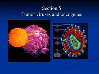
Carcinogenesis and Tumor Viruses. How do viruses contribute to the development of cancer?. Animal Viruses. Lysogeny without the production of infectious virus doesn’t occur in animal cells. Some animal viruses do, however, insert their DNA into the host cell DNA.

Carcinogenesis and Tumor Viruses
E N D
Presentation Transcript
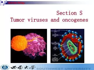
Carcinogenesis and Tumor Viruses How do viruses contribute to the development of cancer?
Animal Viruses • Lysogeny without the production of infectious virus doesn’t occur in animal cells. • Some animal viruses do, however, insert their DNA into the host cell DNA. • Retroviruses – insert their cDNA into random sites in the host cell, but make specific site cuts in the viral cDNA (where are those cuts made?). • This is a necessary step for viral replication. • New viruses are made. • Both horizontal and vertical (germline) transmission of the viruses occur.
Animal Viruses • For DNA viruses, the result of the infection often depends upon whether the host is: • Permissive, in which case a productive, usually lytic infection occurs, or • Nonpermissive, in which case all or part of the viral genome may become inserted into a random site of the host genome. • Cuts in the viral genome are also random. • This is not a useful or necessary step in the replication of the virus.
Animal Viruses • What are some of the effects of the integration of an animal virus on a host cell? • Often the changes that the virus causes in the animal cell make that cell take on the characteristics of a neoplastic or cancer cell. • They don’t respond to the signals or conditions that normally control growth, DNA replication and division. • They become immortal and can grow and divide indefinitely. • This is called neoplastic transformation andviruses that do this are called tumor viruses. • Transformation can be shown by transplanting infected cells, or DNA from transformed cells into a suitable animal host and waiting for the development of a tumor. • Nude mice (have an impaired immune system) may be necessary as the experimental animal (WHY?).
Transformation • Many different morphological and biochemical changes in cultured fibroblasts correlate with transformation: • Cells round up due to cytoskeletal changes • There is a loss of contact inhibition • There is a loss of anchorage dependence • There is a decrease in serum dependence • There are changes in the plasma membrane • There is an increased rate of glycolysis • There is an increased rate of sugar transport
Transformation • Transformation requires continuous expression of only one or a few viral genes. • These genes, therefore, must be very pleiotropic in their actions within the cell. WHY? • This is because cancer is the result a change in cell-cell communication. • Cellular communication begins when a hormone or growth factor binds to a receptor molecule anchored in the plasma membrane (or for non-peptide hormones, diffuses through the plasma membrane and binds to an intracellular receptor).
Transformation • The receptor carries or transduces the chemical signal through the plasma membrane to the internal membrane surface where it activates a second messenger, such as cAMP. • Next a cascade of reactions regulated by protein kinases carries the signal through the cytoplasm and into the nucleus. • In the final step, DNA binding proteins attach to regulatory sequences that initiate DNA replication and/or transcription. • Thus the message to divide or transcribe reaches the genetic material.
Transformation • Transformation always involves an alteration in the regulation of the cell cycle. The cell cycle has four phases: M, during which the cell divides; G1, during which the cell grows larger; S , during which DNA synthesis occurs; and G2, during which the cell continues to grow and prepare for mitosis. The cycle is regulated at several points. • The restrictive point in late G1 phase is a time when the decision is made whether to continue the cycle or to to exit the cycle in a nondividing state called G0. • Cells in G0 may differentiate and assume specialized functions. • A cell can remain in G0 indefinitely, or it may re-enter the cell cycle in response to signals from a variety of growth factors.
Transformation • Once the cell passes the restriction point in G1, the cycle will continue until it is arrested at one of several checkpoints in response to some problem that needs to be corrected. • Progression is halted in late G1 and late G2 if DNA damage has occurred. The checkpoints allow time for the damaged DNA to be repaired before the cycle resumes. • The checkpoint in G2 also responds to the presence of unreplicated DNA and prevents mitosis from occurring until all of the DNA has been copied. • A checkpoint in late M phase halts the cell cycle until all of the chromosomes are properly aligned. • In the event that a cell enters an S phase with damaged DNA, apoptosis may be triggered to prevent the mutant cell from reproducing itself.
The Cell Cycle • Proteins from several different families interact to regulate progression through the cell cycle. • Cyclins, cyclin-dependent kinases (Cdks), and Cdk inhibitors (CKIs) all interact either to block or unblock phases of the cycle. • Cyclins, and Cdks act together as a dimer, functioning as the regulatory and catalytic subunits, respectively. • Cyclins are degraded at the end of their functional period, thus inactivating their Cdk partner in the dimer. • The assembly of the dimers is regulated by other proteins.
An Example of Cell Cycle Regulation by a Serum Growth Factor • Cyclin D is made following the binding of the serum growth factor to its receptor and the ensuing cascade of phosphorylations.
An Example of Cell Cycle Regulation by a Serum Growth Factor
An Example of Cell Cycle Regulation by a Serum Growth Factor • Cyclin D associates with either Cdk4 or Cdk6. • P16 may block the assembly. • After assembly the Cdk becomes phosphorylated. • This may be blocked by either p21 or p27 • The target of the active dimer is Rb which is bound to a transcription factor called E2F. • The Rb/E2F dimer blocks transcription of genes needed to enter the S phase. • Phosphorylation of Rb results in its dissociation from E2F. • This results in activation of S phase genes.
An Example of Cell Cycle Regulation by a Serum Growth Factor • In addition to its ability to block the association of cyclin D with a Cdk, P16 can also directly block the phosphorylation of Rb.
An Example of Cell Cycle Regulation by a Serum Growth Factor • P16, p21, and p27 are regulated by p53 which blocks the cell cycle in the G1 phase if there is DNA damage. • P53, Rb, p21, p16, and p27 are called tumor supressorsbecause their normal function is to prevent the growth of cells with damaged DNA.
An Example of Cell Cycle Regulation by a Serum Growth Factor • P53 also responds to unrepaired DNA damage by triggering apoptosis of the injured cell. • It interacts with a member of the Bcl-2 family of proteins which contains both positive and negative regulators of apoptosis. The Bcl-2 proteins, in turn, activate special enzymes called caspases. • Caspases initiate a protease cascade that results in digestion of the DNA. • This ultimately leads to cell death.
How do Retroviruses Transform Animal Cells? • The genetic information that leads to cellular transformation has no role in the viral reproductive cycle, and in some cases, is not even contained in the genome of the virus. • The basic pattern of the retroviral genome is 5’-gag-pol-env-3’. • Some retroviruses contain another gene that is a tumor causing gene called an oncogene.
How do Retroviruses Transform Animal Cells? • The protein products of the oncogenes have been shown to be necessary and sufficient to cause transformation in cultured cells. • When the sequences of the oncogenes were used as hybridization probes of uninfected cells, it was found that the sequences are in all vertebrate cells.
How do Retroviruses Transform Animal Cells? • This means that the viral gene is actually a cellular gene that became incorporated into the viral genome (remember specialized transduction?) • The cellular gene is called a proto-oncogene. • Retroviruses that contain an oncogene are called transducing viruses.
How do Retroviruses Transform Animal Cells? • Nearly all transducing viruses are replication defective and carry a cellular oncogene in place of the of their own genes. In order to replicate these viruses require a helper virus to supply the missing component(s). • They are called acute transforming retroviruses because they cause transformation shortly after infection.
How do Retroviruses Transform Animal Cells? • Why do oncogenes cause transformation when their cellular counterparts do not? • May be due to quantitative changes in expression (overexpression) • May be due to qualitative differences in expression
How do Retroviruses Transform Animal Cells? • Nontransducing retroviruses don’t carry an oncogene in their genome. They cause transformation months or years after infection. Therefore, they are called chronic transforming retroviruses. • Transformation occurs when the virus inserts next to a cellular oncogene and activates it (via the promoter and/or enhancer sequences of the LTR)
How do Retroviruses Transform Animal Cells? • The activity is cis acting if the activation of the DNA sequence is linked directly to the proviral sequence. • The activity is trans acting if the peptides produced affect activities of other genes that may or may not be linked to the provirus.
What human cancers are caused by retroviruses? • Adult T-cell leukemia - associated with HTLV-I infections • Remember that cancer is multifactorial and the virus infection is only one factor that contributes to the tumor production.
How do DNA Viruses Transform Animal Cells? • DNA viruses that are capable of causing cancer vary from RNA tumor viruses in many aspects: • All families of DS DNA viruses have members that can cause transformation under the right circumstances. • Oncogenic efficiency is usually very low when compared to retroviruses – abortive transformation is common.
How do DNA Viruses Transform Animal Cells? • Small DNA viruses usually only cause transformation when they infect a nonpermissive cell. • Larger DNA viruses may cause transformation in their usual host organism. • The oncogenes that are involved in the transformation are normal viral genes. • Their mechanism of action is as follows:
How do DNA Viruses Transform Animal Cells? • In the nonpermissive cell, early genes are expressed and they force the infected cells into the S phase of the cell cycle. • The cells lack something that allows late gene expression, so there is continuous expression of early genes. • The early gene products directly target the activity of tumor suppressor proteins such as p53 and/or Rb. • If the virus does not integrate this results in abortive transformation. • If the whole virus or part of the virus integrates such that the early genes can be expressed, this may result in stable transformation. • Insertion is mediated solely by cellular enzymes and it is rare and random.
How do DNA Viruses Transform Animal Cells? • Papovaviruses • Polyoma T antigens • Large T antigen is important for initiating transformation, but not for maintaining transformation. • Large T antigen binds to Rb, preventing it from binding to E2F which is then free to activate genes leading to entry into the S phase. • Large T antigen also immortalizes tissue culture cells so that they divide indefinitely and don’t become senescent and die.
How do DNA Viruses Transform Animal Cells? • The middle T antigen is important for the maintenance of transformation by the following mechanism: • Middle T antigen (mT) becomes localized at the plasma membrane and it gets phosphorylated. This is mediated by a phorbal ester. • It activates two cellular protein kinases: • src kinase which phosphorylates mT again. • This creates new binding sites for and activation of phosphatidylinositol kinase (pi3k), phospholipase (Plc ) and the adapter Shc. • Activated pi3k and Plc produce lipids which act as second messengers, relaying signals to various signal transduction pathways. • Activated Shc activates Ras and the Map kinase signal transduction pathway.
How do DNA Viruses Transform Animal Cells? • SV40 T antigens • The large T antigen is multifunctional. In transformed cells it is found in the nucleus where it is responsible for initiation of transformation, and immortalization. • It acts in concert with the small T antigen to maintain the transformation. • Large T antigen exerts its activities in part by binding to and inactivating the growth-regulating functions of Rb and p53.
How do DNA Viruses Transform Animal Cells? • Adenoviruses • E1A and E1B are immediate early genes involved in transformation. • E1A binds to c-ras and to Rb and it transactivates cellular genes involved in the regulation of cell growth. • E1B (55kd) binds to p53 • A second E1B (19kd) protein blocks apoptosis