Download
1 / 1
10 likes | 371 Vues
Materials and Methods:

E N D
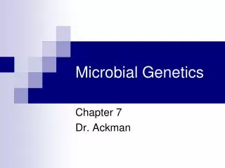
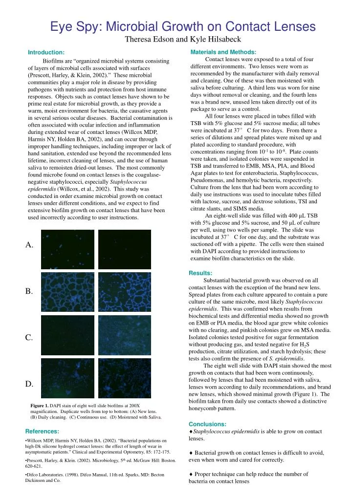
Materials and Methods: • Contact lenses were exposed to a total of four different environments. Two lenses were worn as recommended by the manufacturer with daily removal and cleaning. One of these was then moistened with saliva before culturing. A third lens was worn for nine days without removal or cleaning, and the fourth lens was a brand new, unused lens taken directly out of its package to serve as a control. • All four lenses were placed in tubes filled with TSB with 5% glucose and 5% sucrose media; all tubes were incubated at 37° C for two days. From there a series of dilutions and spread plates were mixed up and plated according to standard procedure, with concentrations ranging from 10-1 to 10-8. Plate counts were taken, and isolated colonies were suspended in TSB and transferred to EMB, MSA, PIA, and Blood Agar plates to test for enterobacteria, Staphylococcus, Pseudomonas, and hemolytic bacteria, respectively. Culture from the lens that had been worn according to daily use instructions was used to inoculate tubes filled with lactose, sucrose, and dextrose solutions, TSI and citrate slants, and SIMS media. • An eight-well slide was filled with 400 μL TSB with 5% glucose and 5% sucrose, and 50 μL of culture per well, using two wells per sample. The slide was incubated at 37° C for one day, and the substrate was suctioned off with a pipette. The cells were then stained with DAPI according to provided instructions to examine biofilm characteristics on the slide. Eye Spy: Microbial Growth on Contact LensesTheresa Edson and Kyle Hilsabeck A. B. C. D. Introduction: Biofilms are “organized microbial systems consisting of layers of microbial cells associated with surfaces (Prescott, Harley, & Klein, 2002).” These microbial communities play a major role in disease by providing pathogens with nutrients and protection from host immune responses. Objects such as contact lenses have shown to be prime real estate for microbial growth, as they provide a warm, moist environment for bacteria, the causative agents in several serious ocular diseases. Bacterial contamination is often associated with ocular infection and inflammation during extended wear of contact lenses (Willcox MDP, Harmis NY, Holden BA, 2002), and can occur through improper handling techniques, including improper or lack of hand sanitation, extended use beyond the recommended lens lifetime, incorrect cleaning of lenses, and the use of human saliva to remoisten dried-out lenses. The most commonly found microbe found on contact lenses is the coagulase-negative staphylococci, especially Staphylococcus epidermidis (Willcox, et al., 2002). This study was conducted in order examine microbial growth on contact lenses under different conditions, and we expect to find extensive biofilm growth on contact lenses that have been used incorrectly according to user instructions. Results: Substantial bacterial growth was observed on all contact lenses with the exception of the brand new lens. Spread plates from each culture appeared to contain a pure culture of the same microbe, most likely Staphylococcus epidermidis. This was confirmed when results from biochemical tests and differential media showed no growth on EMB or PIA media, the blood agar grew white colonies with no clearing, and pinkish colonies grew on MSA media.Isolated colonies tested positive for sugar fermentation without producing gas, and tested negative for H2S production, citrate utilization, and starch hydrolysis; these tests also confirm the presence of S. epidermidis. The eight well slide with DAPI stain showed the most growth on contacts that had been worn continuously, followed by lenses that had been moistened with saliva, lenses worn according to daily recommendations, and brand new lenses, which showed minimal growth (Figure 1). The biofilm taken from daily use contacts showed a distinctive honeycomb pattern. Figure 1. DAPI stain of eight well slide biofilms at 200X magnification. Duplicate wells from top to bottom: (A) New lens. (B) Daily cleaning. (C) Continuous use. (D) Moistened with Saliva. • Conclusions: • Staphylococcus epidermidis is able to grow on contact lenses. • Bacterial growth on contact lenses is difficult to avoid, even when worn and cared for correctly. • Proper technique can help reduce the number of bacteria on contact lenses • References: • Willcox MDP, Harmis NY, Holden BA. (2002). “Bacterial populations on high-Dk silicone hydrogel contact lenses: the effect of length of wear in asymptomatic patients.”Clinical and Experimental Optometry, 85: 172-175. • Prescott, Harley, & Klein. (2002). Microbiology, 5th ed. McGraw Hill: Boston. 620-621. • Difco Laboratories. (1998). Difco Manual, 11th ed. Sparks, MD: Becton Dickinson and Co.