Download
1 / 61
610 likes | 906 Vues
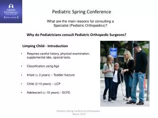
Pediatric Spring Conference. What are the main reasons for consulting a Specialist (Pediatric Orthopedics)?. Why do Pediatricians consult Pediatric Orthopedic Surgeons?. Requires careful history, physical examination, supplemental labs, special tests. Classification using Age

E N D
Pediatric Spring Conference What are the main reasons for consulting a Specialist (Pediatric Orthopedics)? Why do Pediatricians consult Pediatric Orthopedic Surgeons? • Requires careful history, physical examination, supplemental labs, special tests. • Classification using Age • Infant (< 3 years) – Toddler fracture • Child (3-10 years) – LCP • Adolescent (> 10 years) - SCFE Limping Child - Introduction Pediatric Spring Conference Orthopedics - May 8, 2014
Transient Synovitis • Age of presentation – 3-10 years • Presents antalgic gait “refuses to walk” • Normal temperature, low fever, reduced range of motion, mild elevation of ESR • Ultrasound shows effusion of joint – non-echogenic • Lasts 1-3 days • Differential diagnosis • Post streptococcal reactive arthritis – lasts 1-3 weeks • Chronic arthritis – lasts more than 6 weeks Pediatric Spring Conference Orthopedics - May 8, 2014
Infection- Pyarthritis Surgical emergency Irreversible injury to cartilage after 6 hours PredictorProbability Refusal to bear weight 0 predictor 2% Fever greater than 38.5 1 predictor 10% WBC > 12,000 cell/cc 2 predictors 35% ESR > 40 3 predictors 75% CRP > 10 4 predictors > 90% Note – no ultrasound criteria Pediatric Spring Conference Orthopedics - May 8, 2014
Infection- Pyarthritis Organism • Staphylococcus aureus • Streptococcus pneumonia • Group B strep (neonates • Kingella kingae • Treatment • Surgical drainage • Drain placed • IV antibiotics – 3-6 weeks • Osteomyelitis • 1/5000 children less than 13 Pediatric Spring Conference Orthopedics - May 8, 2014
Infection- Osteomyelitis Osteomyelitis Bone involved - Tibia 40% - Femur 20% - Calcaneus 10% - Boy’s 2x’s greater than girls Symptoms • Pain and decreased use 80% • Swelling 60% • Fever 40% Beware of knee pain proximal tibia vs. hip • Blood cultures 50% • Aspiration 80% Pediatric Spring Conference Orthopedics - May 8, 2014
Infection Osteomyelitis Organisms • Staphylococcus aureus 80% • Streptococcus and Gram Negative infancy • Failure of treatment related to polymicrobial infection Diskitis • Triad of symptoms • Pain, fever, reduced intervertebral disc height • Refusal to walk, abdominal pain • Plain x-ray changes – 2 weeks • MRI – most useful • Tx-antibiotics, bracing? Pediatric Spring Conference Orthopedics - May 8, 2014
Hip DDH - After walking age - Painless waddling gait - Unilateral vs. bilateral - Limited abduction, short leg-Galeazzi, Allis - Barlow and Ortolani? After 3-6 months - Plain radiographs - Treatment variable and complicated Pediatric Spring Conference Orthopedics - May 8, 2014
Hip Legg- Calve- Perthes Definition – Ischemia of the proximal femoral epiphysis in children Presentation • 3-12 years old • Boys more common • Painless limp progressing to painful hip • Related to activity • Decreased range of motion Prognosis • Age related • 3-6 years good to excellent • 6-8 years – questionable and variable • > 8 years poor Pediatric Spring Conference Orthopedics - May 8, 2014
Pathology Five Stages • Ischemia • Collapse • Fragmentation • Reconstitution • Remodeling • Investigations • Plain films • MRI • Bone scans – not used much • CT scans – almost never • Labs – none • Radiographic signs • Coxa magna • Coxa vara • Coxa breva • Treatment • Observation • Containment procedures • Procedures to regain strength • Salvage Procedures Hip Pediatric Spring Conference Orthopedics - May 8, 2014
Hip • SCFE • Pathology • Weak interface epiphysis/metaphysis • Common delay in diagnosis • Epiphysis remains with acetabulum Metaphysis displaces anteriorly and superiorly • Most common disorder of adolescent hips • Common in obesity • Ethnicity- • Pacific islanders>African Americans>Caucasian>Asian • Presentation • Adolescent limp • Shortened leg-loss of internal rotation – obligatory external rotation with flexion • Pain – groin-knee-thigh Pediatric Spring Conference Orthopedics - May 8, 2014
Hip • SCFE • Classification • Stable - walking • Unstable – non walking • Prognosis • Depends on: • Stable vs. Unstable • Slip angle - < 45° good - > 45° poor • Treatment • Insitu pinning – single screw center of hip • 2 screws in unstable slips • Open reduction • Hip preservation • FAI • PAO • Osteotomies Pediatric Spring Conference Orthopedics - May 8, 2014
Hip • Chondrolysis • Poorly understood • Limp • Female: Male 5:1 • Age of presentation • Girls - 12.5 years • Boys – 14.8 years • Common delay in diagnosis • Radiographic signs • Decrease in joint space • Acetabular protrusion • Premature physeal obliteration • Marginal osteophytes • Progress is poor with 50% going on to ankylosis usually painless • Associated with SCFE and osteotomies around the hip Pediatric Spring Conference Orthopedics - May 8, 2014
Tibia Toddler Fracture Low energy low impact injury Non displaced or minimally displaced Presents with limp or refusal to walk Investigation • Plain x-ray • Repeat in 2 weeks Treatment Weight bearing cast 3-4 weeks Pediatric Spring Conference Orthopedics - May 8, 2014
Rheumatoid Arthritis Still’s disease Polyarthritic Bimodal age presentation Infants and adolescents-large and small joint involvement. Early diagnosis and medical management. Stiffness – joint swelling – gracile bones – joint deformity Pauciarticular – less painful – swelling <4 joints – Signs, symptoms greater than three weeks. Eye exam is important – Iritis Pediatric Spring Conference Orthopedics - May 8, 2014
Rheumatoid Arthritis Surgical findings Biopsy non-specific inflammatory changes Labs – often not helpful – increased ESR CRP Most other test negative Orthopedics Synovectomy – contracture releases – osteomy – Correction of LLD and total join replacements-arthrodesis Medical management NSAID’s – Cytotoxic agents – Articular steroids – Tumor necrosis block agents – Enbrel – (Etanercept) Pediatric Spring Conference Orthopedics - May 8, 2014
Neoplasms Leukemia Most common cancer in childhood Migratory bone pain ALL most common-presents in infant or child 20% present with musculoskeletal complaints 10% present with a limp Key features Fever, easy bruising, lymphadenopathy and anemia Labs Elevated ESR, thrombocytopenia, anemia, increased lymphoblast's on peripheral smear Radiology Metaphyseal bands – leukemic lines – bone marrow biopsy MRI Pediatric Spring Conference Orthopedics - May 8, 2014
Neoplasms Osteoid Osteoma Benign neoplasm of bone-children or adolescents Classic night pain relieved by NSAID’s 75% of cases to have delayed diagnosis 6 month to 2 years Radiographs – sclerosis with a radiolucent Nidus-bone scan-MRI Surgery Local resection – radiofrequency ablation – Small percentage Resolve spontaneously Pediatric Spring Conference Orthopedics - May 8, 2014
Neuromuscular Disease Pregnancy history, developmental history Screen for cerebral palsy, muscular dystrophy, peripheral neuropathy, spinal dysraphism, spinal muscular dystrophy or ataxia. Most common presenting with a limp is Cerebral palsy with hemiplegia Walking 12-18 months Handedness early Increased gastronemius-soleus muscle tone, upper motor neuron findings – spasticity and hyperreflexia Duchenne Muscular Dystrophy – 2-3 years boys Limp is due to proximal muscle weakness Gower’s sign Elevation of CPK Muscle biopsy Pediatric Spring Conference Orthopedics - May 8, 2014
Knee Discoid Meniscus - Rare – 99% have a lateral meniscus deformity - Most common meniscal disorder in children - Incidence in 1-15% in Japanese and Korean children - Present as snapping or clunking of knee usually associated with minor injuries - Pain effusion and snapping common - Bilateral in 10% - Classification complete, incomplete, Wrisberg - Can have tears of meniscus - Observation – saucerization – subtotal meniscectomy Pediatric Spring Conference Orthopedics - May 8, 2014
Knee Osteochondritis Dissecans - Dissection of bone and cartilage at the ends of long bones - Lateral aspects of medial femoral condyle 70% - Lateral condyle 20% - Patella 10% - Talus - Bilateral 20% - Male to female 1.5:1 - Caused by trauma and subchondral ischemia - Good prognosis in young patients (open growth plates), small lesions (<20mm), no effusion, no dissection on imaging. • Treatment - Observation with activity modification - Drilling non stable lesions - Chondral allograft – small lesions - Chondral autograft – large lesions • Significant sequelae in 20% of patients despite treatment Pediatric Spring Conference Orthopedics - May 8, 2014
Leg Lengths LLD - Painless limp - Toe walking on short leg - Increased knee flexion and hip flexion, hip circumduction on long leg - Hemi hypertrophy/hemi atrophy - Wilms’ tumor – ultrasound Diagnosis • Physical exam • Scanogram • Mild <2 cm Observation • Moderate 2-5 cm Epiphyseodesis • Severe 5-20cm Lengthening • Ridiculous 10-20 cm Combination/Amputation Pediatric Spring Conference Orthopedics - May 8, 2014
Foot Tarsal Coalition - Pain limp - Restricted ROM - Flattening of foot - Most are asymptomatic incidence 1-5% - Bilateral 50% - talocalcaneal, talonavicular - CT scan/MRI - Nonsurgical management most common Surgical coalition resection vs. fusion Diagnosis • Physical examination • MRI inflammation usually mild at insertion points Treatment • Stretching • Limitation of activities • Ice • Bracing/Casting last resort Pediatric Spring Conference Orthopedics - May 8, 2014
- Overuse Apophysistis Presentation Rapidly growing skeleton, increasing body weight, and increased stress due to activity types like sports, very common, non-progressive and usually self-limited. Enthesopathy. Knee Most common site Osgood Schlatter’s disease (OSD) Sinding-Larsen Johansson (SLJ) Foot Achilles Tendon-Severs Disease • Diagnosis • Physical examination • MRI inflammation usually mild at insertion points Treatment • Stretching • Limitation of activities • Ice • Bracing/Casting last resort Pediatric Spring Conference Orthopedics - May 8, 2014
Overuse syndromes in Pediatric Athletes Epidemiology There are 30 million athletes in USA 5-17 years with 4.4 million injuries and 1.4 million serious injuries requiring hospitalization, surgical treatment, missed school and half a day or more of bed rest. Injury rates for Children/100 • Baseball 1.7 • Softball 1.0 • Soccer 2.1 • Football 1.5 Injuries per team per season • Football 14.0 • Baseball 3.0 • Softball 2.0 Pediatric Spring Conference Orthopedics - May 8, 2014
Types of injuries • Contusions most common injuries • Games more common injured than practices • Children have intense training programs, • Vulnerable anatomic regions • (growth plates, joint surfaces, and apophysis) • Gender differences • Sports that involve planting, jumping, and directional change Females have higher injury rate than males • Overall rate for injury rates males to females are 1:2 • ACL injuries in females higher than males • Unique Anatomy • Growth spurts occurs 11 y B , 9 y G • Peak velocities 13.5 boys 11.5 for girls • Growth during puberty comprises 17-18% • Boys double muscle mass during 10-17 years Overuse syndromes in Pediatric Athletes Pediatric Spring Conference Orthopedics - May 8, 2014
Physis • The physis contribute longitudinal growth is susceptible to injury • The region of growth most susceptible to injury is between zone of hypertrophy and calcification • Fractures can be due to acute forces or chronic repetitive forces (stress fractures) • Distal radius – gymnasts • Knee – runners • Ankle/distal fibula – runners • Widening of growth plate • with MRI changes with increased edema Overuse syndromes in Pediatric Athletes Pediatric Spring Conference Orthopedics - May 8, 2014
Apophysis • Rates of skeletal growth exceed rate of growth in muscle therefore relative tension increases • Typical areas affected • Knee – Osgood Schlatter’s disease • Distal Patella • Calcaneus – Severs disease • Medial distal humerus – epicondylitis – little league elbow • Base of 5th metatarsal • Tarsal navicular • Ischial tuberosity • Epiphysis • The epiphysis is another area of vulnerability • Osteochondritis Dissecans – shear forces compression forces, AVN segmental injury • Epiphyseal deformity Overuse syndromes in Pediatric Athletes Pediatric Spring Conference Orthopedics - May 8, 2014
SPECIAL ISSUES IN YOUNG ATHLETES • Intensity, duration and magnitude – key elements that need to be assessed • Approximately 50% of all injuries in sports are overuse • Equal distribution male to female • Older female athletes predisposed to stress fractures and patellofemoral pain • Special Consideration • Shoe type – true running shoes • Proper stretching – 15-30 minutes • Coaching – rapid onset high intensity training • Mechanics – pitching Overuse syndromes in Pediatric Athletes Pediatric Spring Conference Orthopedics - May 8, 2014
Osgood Schlatter’s Disease • Characterized by pain, tenderness and localized swelling of tibial tubercle – clinical exam diagnosis • Reduced activity-application of ice-stretching quadriceps and hamstrings • NSAID’s variable effect • Knee sleeves and straps variable effect • Severs Disease • Characterized by pain, tenderness and localized swelling of Achilles insertion – clinical exam diagnosis – positive squeeze test – soccer • Reduced activity – application of ice – stretching gastrocnemius • NSAID’s variable effect • Heel pads Overuse syndromes in Pediatric Athletes Pediatric Spring Conference Orthopedics - May 8, 2014
Little League elbow and shoulder • Elbow – medial elbow pain, limited extension, significant traction injuries associated with poor throwing mechanics – fewer than 90 pitches, < 200 pitches per week, 3-4 innings per game, mandatory rest periods between games • Shoulder – torsional stresses across shoulder, during cocking and early acceleration phase of throwing, widening of growth plate – Rest – can be severe requiring 3 month no throwing, no curve balls < 14 years • Iselin’s Apophysitis • Pain at the insertion of the peroneus brevis on the proximal fifth metatarsal • Repetitive foot inversion – pain with resisted eversion – NSAID’s, ice, and exercise for increase peroneal strength – casting for severe cases. Overuse syndromes in Pediatric Athletes Pediatric Spring Conference Orthopedics - May 8, 2014
Pelvic Apophysitis • Pain in these specific areas, ages 8-15, active • Iliac Crest • Ischial tuberosity • Anterior superior, iliac apophysis • Rest, ice, NSAID’s, flexibility stretching strengthening • Stress Fracture • mainly in adolescents but can happen in younger children • occur when there are changes in activity levels – intensity • when repetitive force exceeds the reparative process • Locations: • Lumbar spine – gymnasts • Pelvis , femoral neck – dancers-runners • Femoral shaft – Patella, Tibia, Metatarsals, Medial Malleolus, sesamoids – football • Endurance sports most common • Associated eating disorders, amenorrhea, low vitamin D • Tension side injuring more serious than compression side injuries Overuse syndromes in Pediatric Athletes Pediatric Spring Conference Orthopedics - May 8, 2014
Back Injuries • Back injuries are common - 14% incidence • Associated with other diseases such as: • Diskitis, osteomyelitis, and tumors • Disc herniation is less in children than in adults • Dx-MRI, Tx – symptomatic x 6 months • Spondylolysis has a 6% incidence most commonly Asymptomatic and if painful conservative measure likely Effective rarely bracing is required • Gymnastics, dance, and diving • Diagnosis • Plain X-ray • CT scan • MRI • Congenital and acquired • Treatment • Symptomatic • Bracing • Surgery • Can be associated with spondylolisthesis Overuse syndromes in Pediatric Athletes Pediatric Spring Conference Orthopedics - May 8, 2014
Overuse syndromes in Pediatric Athletes • Strength Training • Recommend against in children and preadolescents • Adolescent strength training programs can reduce injuries • 300 soccer players’ 14-18 years. • 7 –week preseason plyometric exercises reduced injuries from 33%-14% • Males training 0.09% injuries – untrained females 4.8 times higher Pediatric Spring Conference Orthopedics - May 8, 2014
Pediatric Orthopedic Infections Osteomyelitis • 1 in 5000 children younger than 13 years • More common in first decade • Boys 2.5 times more common • Pre-modern era mortality 50% - modern era mortality 1% Acute Hematogenous Osteomyelitis • Most common presentation • Femur and tibia most commonly affected • Risk factors: • Diabetes • Hemoglobinopathies • Chronic renal disease • Rheumatoid arthritis • Immunosuppression Pediatric Spring Conference Orthopedics - May 8, 2014
Pediatric Orthopedic Infections Pathogenesis Trauma => slower flow end arteries metaphyseal bone =>bacterial showers seed region – Cellulitic phase can be treated with antibiotics Purulence extends through porous bone and penetrated cortex Elevated periosteum – bone necrosis Sequestrum is loosely adherent dead bone Involucrum is new bone over dead bone – can Lead to septic arthritis in hip and shoulder Lab WBC- elevated in only 25% ESR - elevated in 90% peaks at 3-5 days, declines in 1-2 weeks CRP – elevated in 98% rapid rise in 24-48 hours decrease within 6 hours of antibiotics Blood cultures – positive in 30-36% Pediatric Spring Conference Orthopedics - May 8, 2014
Pediatric Orthopedic Infections Imaging Plain films MRI gold standard Bone scans rarely done Ultrasound limited value Organisms Staphylococcus aureus most common – 60-90% Group A strep Group B strep – neonate H flu nearly wiped out due to vaccination Salmonella consider sickle cell disease 3-10% MRSA Special note Neonates Consider multiple sites up to 40% Lab tests are not helpful Recurrent infection is primary presentation of resistance due to multiple site infections Pediatric Spring Conference Orthopedics - May 8, 2014
Septic Arthritis Surgical Emergency • Particular issues with hip and shoulder due to vascular anatomy – AVN and septic physeal separation • Proteolytic enzyme release causing damage to articular cartilage • Mostly Staphylococcus aureus • Joints involved: • Hip 35% • Knee 35% • Ankle 10% • Wrist, Elbow, Shoulder 15% • Small joints 2% Organisms • S Aureus 56% • Group A strep 22% • S Pneumonia 6% • Other gram negatives Klebsiellae, salmonellae, Kingella • H. influenzae B 40% pre vaccination Pediatric Spring Conference Orthopedics - May 8, 2014
Differential Diagnosis • Culture negative disease Gonococcal • Osteomyelitis • Lyme disease • TB • Transient synovitis • Post strep reactive • Henoch Shonlein purpura • Hemophilia • Sickle cell disease • Leukemia • PVNS • Etc. Labs • CRP, ESR,CBC, Differential, Blood culture, throat swabs, rapid strep test, Antistreptolysin O titer, Lyme disease titer • Blood cultures positive 30-36% • Infectious organism identified in 50-70% • Elevated WBC 30-60% • Left shift 60% • Positive result of treatment • CRP < 20 in 7 days • ESR < 25 in 3 weeks • Aspiration of joint • Clinical and lab values indicate infection • Hip and should differentiate open arthrotomy from no arthrotomy equivocal clinical and labs • When in doubt open joint and do formal washout with drain • Labs – CBC cultures aerobic anaerobic fungal TB, fluid glucose, and Gram stain • > 25,000 wbc • < 0.5 fluid to serum glucose Pediatric Spring Conference Orthopedics - May 8, 2014
Other infections Diskitis and Vertebral Osteomyelitis Diskitis • Uncommon in children • Lumbar region in children younger than 5 years • Present with refusal to walk + Gower like sign • Low-grade fever • Disc narrowing on lateral plain film • MRI is gold standard look for paravertebral infection and osteomyelitis of contiguous vertebrae • Treatment antibiotics alone most commonly Vertebral Osteomyelitis • Older children • Present with back pain focal tenderness • More febrile appear more ill than Diskitis • Aspiration biopsy • S. Aureus most common • Antibiotics alone commonly effective • Lack of response may require surgery Illiopsoas Abscess • In the differential of septic arthritis of the hip • Passive rotation of the hip is painless • Diagnosis – CT, MRI, Ultrasound • Treatment – percutaneous drainage procedure Pediatric Spring Conference Orthopedics - May 8, 2014
Lyme Disease • Multisystem disease with spirochete B burgdorferi • Transmitted through bite of infected lxodes tick • Northeast USA • May to August • Tick attachment > 24 hours • Skin lesion erythema migrans – bull’s-eye appearance • Symptoms • Fever • Headache • Malaise • Arthralgias • Multiple secondary skin lesions • Cardiac a-v blocks • Arthritis • Encephalopathy • Polyneuropathy • Knee is most common joint involved • Oral doxycycline 3-4 weeks 90% effective • IGG test takes 4-6 weeks Pediatric Spring Conference Orthopedics - May 8, 2014
Musculoskeletal Neoplasm Assessment History • Pain at night or rest • Pain for more than 6 weeks • Mass • Age • Location • Duration Physical Examination • Lymph node regions • Lung • Mass size and mobile of fixed • Deep or superficial • ROM around joints • Muscle atrophy, angular deformity, LLD • Single site multiple sites Pediatric Spring Conference Orthopedics - May 8, 2014
Diagnostic Imaging • Plain films most important • Four questions: • What is location of lesion? – Age of patient? • What is the tumor doing to the surrounding bone? • What is the bone doing in response to the tumor? • What is the matrix of the tumor? • CT Scan • MRI Scan Pediatric Spring Conference Orthopedics - May 8, 2014
Neoplasm Staging • Benign • Latent • Active • Aggressive Malignant • l Low grade • II High grade • III Metastatic Principles of Biopsy • Don’t do it unless you are the surgeon going to do definitive procedure • Longitudinal incisions • Stay away from neurovascular structures • No planar dissection • Stay in one compartment • Meticulous hemostasis • Drain in line with incision • Subcuticular closure • Compression dressing • Incisional biopsy Pediatric Spring Conference Orthopedics - May 8, 2014
Osteoid Osteoma Bone Tumors by Tissue Type Bone Osteoblastoma Pediatric Spring Conference Orthopedics - May 8, 2014
Osteoblastoma Cartilage Enchondroma Pediatric Spring Conference Orthopedics - May 8, 2014
Oillier’s Disease Osteochondroma Pediatric Spring Conference Orthopedics - May 8, 2014
Hereditary Multiple Exostoses Chondroblastoma Fibrous Fibrous Dysplasia Pediatric Spring Conference Orthopedics - May 8, 2014
Osteofibrous Dysplasia Nonossifying Fibroma Pediatric Spring Conference Orthopedics - May 8, 2014
Miscellaneous Ewing's Sarcoma Giant Cell Tumor Pediatric Spring Conference Orthopedics - May 8, 2014
Unicameral Bone Cyst Aneurysmal Bone Cyst Langerhans Histiocytosis Pediatric Spring Conference Orthopedics - May 8, 2014