Download
1 / 31
310 likes | 459 Vues
This study presents innovative 3D multimodal visualization techniques designed to enhance tumor resection surgeries in neurosurgery. It emphasizes the integration of intraoperative MRI with preoperative data to improve surgical planning and real-time navigation. Key advancements include the development of a new multi-volume rendering algorithm based on depth peeling, which allows surgeons to visualize critical structures such as blood vessels and functional brain areas, thereby reducing occlusions and enhancing the clarity of anatomical context during high-risk procedures.

E N D
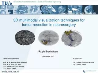
3D multimodal visualization techniques for tumor resection in neurosurgery Ralph Brecheisen 18 december 2007 Graduation committee: Prof. dr. ir. Bart ter Haar Romeny Prof. dr. ir. Jack van Wijk Dr. ir. Anna Vilanova i Bartroli Dr. ir. Bram Platel Supervisors: Dr. ir. Anna Vilanova i Bartroli Dr. ir. Bram Platel
Introduction Department of Neurosurgery azM Image-guided Surgery TU/e (Dr. Bram Platel) Intraoperative navigation Enhance i-MRI with preoperative data Resection verification Polestar N20 i-MRI Medtronic StealthStation Tumor resections Improve surgery planning
Brain tumor resections Functional activations High-risk Procedures Blood vessels Fiber tracts Tumor Image: Yale University
Investigate requirements Blood vessels Functional activations Fiber tracts Tumor Anatomical context ? - 2-dimensional - Not spatially related Too difficult! Images: University of Florida, Music Therapy World, King Edward Memorial Hospital, Neuro Imaging UCLA, National Institutes of Health
What do we want to do? 3D! Blood vessels Functional activations Fiber tracts Anatomical context Tumor Combine in single visualization Images: university hospital Maastricht, Stanford University, M. Meissner Viatronix Inc
What else do we need? Reduce occlusions and clutter Intraoperative navigation Hide/show Surgical tools 3D pointers Grid lines Cut outs (clipping) Geometric models Semi- transparency
How to deal with the volume data? • Reviewed existing implementations • Main points: • Data representation • Rendering
Data representation B Different scan times Interpolation inaccuracies Different volume of interest Memory waste A Resampling Intraoperative data
Rendering Ray casting Texture slicing screen screen D D D • - Adaptive sampling • Programmable GPU • Works for old GPU’s-Not very flexible
Review conclusion • Nobody does combination of • Ray casting • Render without resampling • Intersecting semi-transparent geometry How do WE render multiple volumes?
y x z How to render a single volume? Viewing window • Vertex • Color • Position 2D projection Polygon Bounding box
Rasterization I • Fragment • Color • Depth • ScreenCoord (i,j) j Dfrag Cube Viewing window (0,0) i Screen
Rasterization II Default: depth test LESS nearest fragments written to screen Other depth tests: EQUAL, GREATER
How to link bounding box with dataset? Vertex (1,1,1) (0,0,0) 3D texture = dataset (in GPU memory) • Fragment • Color/depth/screenpos • Interpolated texture coordinate
Programmable fragment shaders User-defined parameters - E.g. texture ID’s Fragment shader Updated color Fragment Tex ID Looping Boolean logic, math functions Random texture lookup
v v* Hardware-accelerated ray casting Fragment shader: Take sequence of texture samples along viewing direction Mix samples Screen frag Projection image 3D texture Nearest fragments
How to mix texture samples? opacity bone 1 skin 0 Transfer function image courtesy: Washington University Medical Center α1 α2 α3 … Color samples Blending equation
How could we render multiple volumes? Check position at every step! Use single pass Screen B P P’ P’’ A 3D Texture 1 3D Texture 2
Multi-volume depth peeling Nearest fragments Second-nearest fragments Subdivide volumes in regions DEPTH LAYERS Screen 2D accumulation texture CACC
Initialization 2D textures Screen DNEAR CNEAR CACC = (0,0,0,0) Depth test LESS
if(Dfrag≤ Dnear ) then Discard() endif Depth peeling I Screen second-nearest fragments depth test LESS Peeling shader DFAR DNEAR CFAR
Ray casting I depth test EQUALDNEAR DNEAR Raycasting shader: (1) Compute start/end positions in region (2) Take Cacc (3) Blend with Cnear (4) Blend with texture samples (5) Output color DFAR CACC CACC CNEAR
DNEAR DFAR CNEAR CFAR if(Dfrag≤ Dnear ) then Discard() endif Depth peeling II Screen Swap textures third-nearest fragments Peeling shader DFAR DNEAR CFAR
Ray casting II depth test EQUALDNEAR Screen DNEAR DFAR Raycast shader CACC CACC CNEAR Screen
Intersecting geometric models Almost for Free! Screen Sphere Volume bounding box Raycasting shader: … (2) Take Cacc (3) Blend with Cnear … Cnear = blue Cnear = 0,0,0,0
Cut outs (clipping) Clipping sphere Screen DFAR DNEAR
Results A picture says… DEMO!
Performance NVIDIA GeForce 8800 GTX, 768MB (screen: 500x500)
Conclusions • Investigated surgery planning requirements • Developed new multi-volume rendering algorithm based on depth peeling • N volumes, N transfer functions • Translucent geometry intersections • Convex clipping • Interactive displacement of volumes and geometry • Basic user interface
Future work • Build neurosurgery application • Integrate with i-MRI and navigation system • Combine gray value TF’s with object segmentation • Integrate DTI tool Anna • Enhance structures using multimodal info • Memory handling and performance