Download

1 / 110
1.1k likes | 1.32k Vues
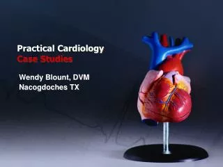
Practical Cardiology Case Studies. Wendy Blount, DVM Nacogdoches TX. Daisy. Signalment 15 year old spayed female mixed terrier 11 pounds Chief Complaint Became dyspneic while on vacation, as they drove over a mountain pass

E N D
Practical CardiologyCase Studies Wendy Blount, DVM Nacogdoches TX
Daisy Signalment • 15 year old spayed female mixed terrier • 11 pounds Chief Complaint • Became dyspneic while on vacation, as they drove over a mountain pass • Come to think of it, she has been breathing hard at night for some time
Daisy Exam • T 100.2, P 185, R – 66, BP – 145, BCS – 3.5 • Increased respiratory effort (heart sounds) • 3/6 holosystolic murmur loudest at left apex • Mucous membranes pale pink • Crackles in the small airways • Pulses weak, somewhat irregular, no pulse deficits • CRT 3.5-4 seconds
Daisy Differential Diagnosis - Dyspnea • Suspect congestive heart failure • Suspect mitral regurgitation • Concurrent respiratory disease can’t be ruled out Initial Diagnostic Plan • Chest x-rays, ECG • CBC, mini-panel, electrolytes
Daisy CBC, mini-panel, electrolytes • Normal
Daisy CBC, mini-panel, electrolytes • Normal
Daisy CBC, mini-panel, electrolytes • Normal
Daisy CBC, mini-panel, electrolytes • Normal Thoracic radiographs • Markedly enlarged LA • Compressed left mainstem bronchus • Perihilar edema • Vertebral heart score 11.75 • Elevated trachea – LV enlargement • Right heart enlargement, enlarged pulmonary lobar aa. • Mildly enlarged liver • Enlarged caudal vena cava
Daisy ECG
Daisy Calculating Instantaneous Heart Rate (iHR) • Measure R wave to R wave (9mm) • Divide by paper speed (25 mm/sec) for time per beat 9mm x _sec_ = 0.36 sec per heart beat 25mm • Calculate beats per minute _heart beat_ x _60 sec = 166 beats/minute 0.36 sec minute
Daisy ECG • Rate – 110 bpm • Rhythm – sinus arrhythmia with VPCs • MEA – normal (lead II has tallest R waves) • P, QRS and T waves – normal • No evidence of enlarged LA and LV on the ECG • VPC – abnormal QRS • Comes too early (166 bpm) • Wide and bizarre shape • Not preceded by a P wave • T wave opposite in polarity than normal QRS
Daisy Initial Therapeutic Plan • Lasix 25 mg IM, then 12.5 mg PO BID • Enalapril 2.5 mg PO BID • Owner is a lab tech, and set up oxygen mask to use PRN at home • Recheck BUN, potassium, chest rads 3-5 days • Come back sooner if respiratory rate at rest is above 40 per minute without oxygen
Daisy When to treat VPCs • VPCs unusual for MR • Did not treat in this case, because: • MR dogs not predisposed to sudden death • SAS and DCM are most common causes of sudden death due to arrhythmia • Ectopic focus not firing at a fast rate (166 bpm) • <200 bpm iHR is well away from the T wave • No pulse deficits – did not affect hemodynamics • Primary treatments for VPC are Sotalol or B blocker • Negative inotropes not ideal for myocardial failure
Daisy Recheck – 4 days • Daisy’s breathing is much improved (30-40 at rest) • Lateral chest x-ray • Electrolytes normal • BUN 52
Daisy Recheck – 4 days • Daisy’s breathing is much improved (30-40 at rest) • Lateral chest x-ray • Electrolytes normal • BUN 52
Daisy Diagnostic Plan - updated • Decrease enalapril to SID • Recheck BUN 1 week • Recheck chest rads 1 week Recheck – 1 week • BUN – 37 • Thoracic rads no change • Request recheck in 3 months, or sooner if respiratory rate at rest is above 40 per minute
Daisy 2 months later • Daisy is breathing hard again at night Exam • Same as initial presentation Diagnostic Plan • CBC, mini-panel, electrolytes • Chest x-rays
Daisy 2 months later • Daisy is breathing hard again at night Exam • Same as initial presentation Diagnostic Plan • CBC, mini-panel, electrolytes • Chest x-rays
Daisy 2 months later • Daisy is breathing hard again at night Exam • Same as initial presentation Diagnostic Plan • CBC, mini-panel, electrolytes • Chest x-rays
Daisy Bloodwork • CBC, electrolytes normal • BUN 88 Therapeutic Plan • Increase furosemide to 18.75 mg PO BID • Add hydralazine 2.5 mg PO BID • Recheck chest rads, BUN, electrolytes, blood pressure 1 week
Daisy Recheck – 1 week • Clinically much improved – respiratory rate 30-40 per minute at rest • electrolytes normal • BUN 58 • Blood pressure 135 • Chest x-rays • Recommend recheck in 3 months, or sooner if respiratory rate above 40 per minute at rest
Daisy Recheck – 1 week • Clinically much improved – respiratory rate 30-40 per minute at rest • electrolytes normal • BUN 58 • Blood pressure 135 • Chest x-rays • Recommend recheck in 3 months, or sooner if respiratory rate above 40 per minute at rest
Daisy Recheck – 6 months • Daisy dyspneic again Exam • Similar to last crisis – BP 90 Diagnostic Plan • CBC, mini-panel, electrolytes • Echocardiogram, ECG, chest x-rays
Daisy Bloodwork • CBC, electrolytes normal • BUN 105, creat 2.1 Chest x-rays
Daisy Bloodwork • CBC, electrolytes normal • BUN 105, creat 2.1 Chest x-rays
Daisy Bloodwork • CBC, electrolytes normal • BUN 105, creat 2.1 Chest x-rays • Similar to last crisis ECG • Sinus tachycardia, wide P wave
Daisy - Echo Short Axis – LV apex (video) • LV looks big Short Axis – LV papillary muscles • IVSTD – 6.0 mm – low normal • LVIDD – 35 mm (n 20.2-25) • LVPWD – 4.3 mm – low normal • IVSTS – 9.4 mm – normal • LVIDS – 25 mm (n 11.1-14.6) • LVPWS – 8.4 mm - normal
Daisy - Echo Short Axis – LV papillary muscles • IVSTD – 6.0 mm – low normal • LVIDD – 35 mm (n 20.2-25) • LVPWD – 4.3 mm – low normal • IVSTS – 9.4 mm – normal • LVIDS – 25 mm (n 11.1-14.6) • LVPWS – 8.4 mm – normal • FS – (35-25)/35 = 29% (normal 30-46%)
Daisy - Echo Short Axis - MV • MV leaflets hyperechoic and thickened • EPSS – 8 mm (n 0-6) Short Axis – Aortic Valve/RVOT • LA appears 2-3x normal size • AoS – 13.0 – normal • LAD – 33 mm (n 12.8-15.6) • LA/Ao = 2.5 (n 0.8-1.3)
Daisy - Echo Long View – 4 Chamber • LV and LA both appear large • MV is very thick and knobby, with some prolapse into the LA
Daisy - Echo Long View – 4 Chamber • LV and LA both appear large • MV is very thick and knobby, with some prolapse into the LA
Daisy - Echo Long View – 4 Chamber • LV and LA both appear large • MV is very thick and knobby, with some prolapse into the LA • Pulmonary vein markedly enlarged Long View – LVOT • Large LA, Large LV (video)
Daisy Therapeutic Plan • Increase hydralazine to 5 mg PO BID • Add spironolactone 12.5 mg PO BID • Add pimobendan 1.25 mg PO BID • Increase furosemide to 18.75 mg PO TID x 2 days, then decrease to BID if respiratory rate decreases to less than 40 per minute at rest. • Recheck 1 week – BUN, creat, phos, electrolytes, chest rads, BP
Daisy Recheck – 1 week • Clinically improved again • BP - 125 • BUN 132, creat 2.6, phos 6.6 • Electrolytes normal • chest rads improved pulmonary edema Therapeutic Plan – Update • Add aluminum hydroxide gel 2 cc PO BID
Daisy 5 Months later • Coughing getting worse • Chest rad show no pulmonary edema • LA getting larger Therapeutic Plan – Update • Add torbutrol 2.5 mg PO PRN to control cough
Daisy 18 Months after initial presentation • Owner discontinue pimobendan due to GI upset 28 months after initial presentation • Daisy finally took her final breath • BUN >100 for 22 months
Chronic MV Disease • May be accompanied by similar TV disease (80%) • TV disease without MV disease possible but rare • LHF and/or RHF can result • Right heart enlargement can develop due to pulmonary hypertension, in turn due to LHF • Myocardial failure and CHF are not directly related
Chronic MV Disease Thoracic radiograph abnormalities: • LV enlargement • Elevated trachea • increased VHS • LA enlargement – often largest chamber • Compressed left bronchus • + left heart failure • Pulmonary edema • Lobar veins larger than arteries
Chronic MV Disease Echo abnormalities: (doppler echo) • LA and/or RA dilation, LV and/or RV dilation • Exaggerated IVS motion (toward RV in diastole) • Increased FS first, then later decreased FS • Thickened valve leaflets • If TV only affected, left heart can appear compressed, small and perhaps artifactually thick • Ruptured CT – • MV flips around in diastole • MV flies up into LA during systole – “MV flail” (video) • May see trailing CT, or CT floating in the LV
Chronic MV Disease ECG abnormalities: • Wide or notched P wave • Enlarged LA • Tall R wave • Enlarged LV • Right Bundle Branch block • Wide QRS • Deep S wave • Left Bundle Branch Block • Wide QRS • Tall R wave
Chronic MV Disease Right Heart Failure • Medications similar to LHF • Medications not as effective at eliminating fluid congestion • More effective at preventing fluid accumulation, once controlled • Periodic abdominocentesis and/or pleurocentesis required • Prognosis for RHF and LHF is extremely variable
Chronic MV Disease Classification of Chronic AV Valve Disease • Class I - small, discrete nodules along the edge of the valve leaflets • Class II - free edges are thickened and the edges of the leaflets become irregular. Some CT are thickened. • Class III - valve edges grossly thickened and nodular, extending to the base of the valve leaflets. There is redundant tissue, resulting in prolapse into the LA. CT are thickened and may rupture, resulting in mitral valve flail. CT to the septal leaflet can also elongate.
Chronic MV Disease LA Jet Lesions • fibrous plaques in the endocardium in a region subjected to the impact of the high velocity MR jet. • Endomyocardial splits or tears may also be identified. • On occasion, a full thickness left atrial tear occurs resulting in hemopericardium, pericardial tamponade, and usually death. • Rarely, a full thickness endomyocardial tear will involve the interatrial septum, causing an acquired atrial septal defect. (MR Client Handout)
MVD in Cavaliers • Leading cause of death in Cavaliers • CHF can develop as young as 1-3 years old • First sign of disease is mitral murmur • Careful annual auscultation • Radiographs should be done as soon as murmur is detected • q6months when progressing • annually for stable disease • Sooner when respiratory rate exceeds 40 per minute • Doppler Echo when abnormalities are present on rads
MVD in Cavaliers • The median survival period from grade III CHF due to MVD is approximately seven months, with 75% of the dogs dead by one year • Current recommendation is that no Cavalier be bred until after 5 years of age, with no murmur • At this time, a majority of Cavaliers are affected • Many progress to grade II CHF (Client Handout)
Susie Signalment • 12 year old spayed miniature schnauzer Chief Complaint • Episodes of Confusion Exam • G3 dental tartar • Alternating periods of normal heart rate, tachycardia and bradycardia • Pulse deficits during tachycardia
Susie Work-up • CBC, panel, electrolytes, UA normal • Chest x-rays
Susie Work-up • CBC, panel, electrolytes, UA normal • Chest x-rays Vertebral Heart Size = 10.7 (normal 8.5-10.5) Enlarged main pulmonary artery
Susie Work-up • CBC, panel, electrolytes, UA normal • Chest x-rays • Susie is not on heartworm prevention
Susie Work-up • CBC, panel, electrolytes, UA normal • Chest x-rays • Susie is not on heartworm prevention