
Introduction
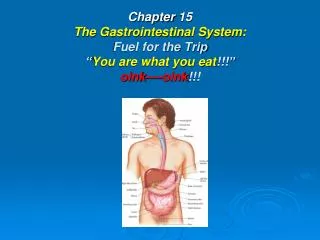
Chapter 15 The Gastrointestinal System: Fuel for the Trip “ You are what you eat !!!” oink----oink !!!. Introduction. Gastrointestinal System Functions: Ingest raw materials Physically & chemically digest raw material to usable elements. Absorb elements Eliminate what is NOT useable.

Introduction
E N D
Presentation Transcript
Chapter 15The Gastrointestinal System:Fuel for the Trip“You are what you eat!!!”oink----oink!!!
Introduction Gastrointestinal System Functions: • Ingest raw materials • Physically & chemically digest raw material to usable elements. • Absorb elements • Eliminate what is NOT useable
System Functions • Ingestion: food enters mouth • Mastication: chewing • Digestion: chemical act of breaking down food into small molecules. • Secretion: acids, buffers, enzymes, & H2O aid in breakdown of food. • Absorption: molecules pass through lining of digestive tract. • Excretion or defecation: elimination of waste products.
Buccal (oral) Cavity • Lips: act as door to cavity • Hard & Soft Palate: form roof of mouth • Tongue: acts as floor • Cheeks: form walls
Tongue • Muscle: provides taste stimuli to brain, determines temperature, manipulates food, & aids in swallowing. • Saliva: added to moisten & soften food, while teeth crush food. • Bolus: ball-like mass, pushed by tongue so it may be swallowed, passed to pharynx. • Lingual Frenulum: membrane under tongue, keeps you from swallowing tongue & aids in speaking.
Salivary Glands • Sublingual: found under tongue • Submandibular: located along both sides of inner surface of mandible, or lower jaw. • Controlled by: autonomic nervous system • Parotid: slightly inferior & anterior to each ear. Swell with “viral Parotitis..”
Salivary Glands con’t • Produces: 1–1.5 liters of saliva QD • Keep mouth moist: but idea or presence of food increase production significantly. • Contains: 99.4% water, & contains antibodies, buffers, ions, waste products, & enzymes.
Salivary Glands con’t • Enzymes: act as organic catalysts to speed up chemical reactions. • Salivary Amylase: speeds chemical activity of breaking down carbohydrates. • Saliva cleans oral surfaces, reducing amount of bacteria that grows in mouth.
Teeth • Deciduous: first set of teeth as a baby • First tooth: appears @ 6 months of age; lower central incisors appear first, all 20 teeth in place by age 2½. • Between 6 and 12 years these teeth fall out, are replaced by 32 permanent teeth. • Wisdom teeth: appear by age 21
Teeth Con’t • Incisors: at front of mouth, blade shaped, used to cut food. • Canine: for holding, tearing, or slashing food; known as eyeteeth or cuspids, located next to incisors. • Bicuspids: or premolars: transitional teeth • Molars: have flattened tops; both bicuspids & molars are responsible for crushing & grinding food.
Parts of Tooth: Crown: covered by hard enamel. Neck: transitional section that leads to root. Root: nestled in bony socket, held in place by fibers of periodontal ligament. Dentin: made of mineralized bone-like substance. Teeth Con’t
Connective tissue: pulp, located in pulp cavity Pulp cavity: contains blood vessels & nerves providing nutrients & sensation; nerves & blood vessels get to pulp cavity via root canal. Cementum: (soft version of bone) covers dentin of root, aiding in securing periodontal ligament. Teeth Con’t
TeethCon’t • Gingiva: gums, help hold teeth in place • Epitheal cells form tight seal around tooth to prevent bacteria from coming into contact with tooth’s cementum.
Pathology Connection: Oral Disorders Dental Caries (cavities) • Form whenmicroorganisms attack tooth enamel • Related to dental plaque: sticks to teeth forming sticky substance. • Forms great hideout for bacteria • Bacteria createsacids that attack surface of teeth.
Risk Factors for Plaque Formation • High carbohydrate diet • Poor dental hygiene • Lack of regular visits to dentist
Risk Factors for Plaque Formation con’t RX: • Clear out & fill caries • Rx infection Prevention: • Proper dental care • Fluoride in H2O & tooth paste • Evaluate for heart disease & buccal ca
Pathology Connection:Periodontal Disease • Plaque & bacteria affects gums & supportive structures of teeth. • Can result ingingivitis, bleeding & tooth loss
Pathology Connection:Oral & Lip Cancer Cause: • Excessive sun exposure • Tobacco • ETOH
Oral & Lip Cancer con’t Lukoplakia: • white patch of tissue in mouth • associated with use of chewing tobacco
Pathology Connection:Stomatitis • Inflammation of oral mucosa • poor fitting dentures • Apthousstomatitis (“canker sores”) • Cheilitis: cracking & inflammation of lips & corners of mouth; often related to infection, allergy, or nutritional deficiency.
Pharynx (3 Parts) Nasopharynx: • primarily part of respiratory system, blocked by soft palate. Oropharynx & laryngopharynx: • act as passageway for food, water, & air; epiglottis covers trachea to prevent food from entering lungs, forcing food into opening for esophagus.
Esophagus • 10 inches long, is connected to stomach • from pharynx, through thoracic cavity, through diaphragm, connecting to stomach in peritoneal cavity. • normally collapsed tube until “bolus” of food swallowed. • Peristalsis: pushes food down esophagus
Esophagus con’t • lined withstratified squamous epithelium that secrete mucus to make walls slippery; cells make lining. • resistant toabrasion, temperature extremes, & irritation. • Pharyngoesophageal sphincter: relaxes to open esophagus so food can enter.
Esophagus con’t • Lower Esophageal Sphincter: opening door to stomach & closing to prevent acidic gastric juices from splashing into esophagus causing heartburn. • process of swallowing: food9 seconds; fluid take only seconds to reach stomach.
Located: ULQ under diaphragm, posterior to Liver. 10 inches long with diameter dependent on how much just eaten. 4 liters when filled Rugae: folds, help stomach expand and contract. Stomach
4 Function of Stomach • Holdingarea for received food • Chemical digestion: gastric acids & enzymes mix with food. • Regulates rate of Chyme movement into small intestines. • Absorbs small amounts of H2O & ETOH
How Fast Stomach Empties • 4 hours to empty following meal • Liquids & carbohydrates pass quickly • Proteins take longer • Fats take longest 4-6 hours
Cardiac Region: surrounding lower esophageal sphincter. Fundus: laterally & slightly superior to cardiac region. Temporarily holds food as it enters stomach. Body: Mid-portion Pylorus: 1. terminal end of stomach 2. most of work performed 3. where food passes through pyloric sphincter into small intestine. 4 Regions of Stomach
Chemical Digestion Gastric Juice: • 1500 mls produced QD • hydrochloric acid (HCl) • pepsinogen • mucus
Pepsinogen, HCL & Pepsin Enzymes • chief digestive enzyme • secreted bychief cells • HCL secreted by parietal cells combining to produce pepsin. • Pepsin breaks down protein • HCl breaks down connective tissue
Stomach & Enzymes con’t • HCL: pH of 1.5–2.0, effective at killing pathogens. • Mucous cells: generate thick layer of mucus shielding stomach from effects of stomach acids. • Stomach secretes intrinsic factor, allowing vitamin B12 to be absorbed. • Enzyme activity controlled by parasympathetic nervous system (vagus nerve) • Vagus increases motility & secretory rates of gastric glands.
I. Cephalic Phase: sensory stimulation (sight or smell of food) stimulates parasympathetic nerves via medulla oblongata Gastrin released stimulating gastric gland activity in stomach 3 Phases of Gastric Juice Production
3 Phases of Gastric Juice Production con’t II. Gastric Phase: • 2/3 of gastric juices secreted as food enters stomach & distends walls. • signaling stomach to secrete more gastric fluid
3 Phases of Gastric Juice Production con’t III. Intestinal Phase: • food enters duodenum, distending & sensing acidity. • intestinal hormones released • slowing gastric gland secretions • lasts until bolus leaves duodenum
Rate of Movement of Chyme If too slow: • rate of nutrient digestion & absorption decreased • may allow acidity of chyme to cause erosions of stomach lining (ulcers). If too quick: • food particles may not be sufficiently mixed with gastric juices. • insufficient digestion; chyme not given time to neutralize can cause erosion of intestinal lining (ulcers).
Pathology Connection: Stomach Acid Disorders • Gastroesophageal Reflux Disease (GERD) • Condition where acidic stomach contents “squirt” back into esophagus • Since esophagus does not have protective mucus, can cause inflammation and ulceration of esophageal tissue • Scar tissue can eventually form, causing narrowing of esophagus • If left untreated, constant inflammation can lead to esophageal cancer
GERD cont. • s/sepigastric pain and burning, can be worse when lying down • d/x symptoms, upper GI • R/x • Antacids: treat burning sensation by decreasing acid • Acid reducing meds • Lifestyle changes: may help prevent GERD • Limiting fats, alcohol, caffeine and chocolate in diet • Avoiding smoking • Avoiding lying down in 4 hours after eating • Sleeping with head of bed elevated • If obese, weight loss
Peptic Ulcers Etiology: Breakdown of mucosal membrane in esophagus, stomach, or small intestine; develop most commonly in duodenum Factors that increase risk: • Helicobacter pylori (H. pylori) infection in stomach: • Smoking • Heavy/chronic alcohol consumption • Use of NSAID medications (including aspirin and others) • Caffeine consumption
Peptic Ulcer cont. • Use of corticosteroid medications • Stress
Small Intestine • major organ of digestion, is where most of food digested • average length of 6–20 feet and diameter ranging from 2.5-4 cm • Walls secrete digestive enzymes and hormones to stimulate pancreas
Small Intestine cont. • 80% of absorption of usable nutrients occurs in sm. Intestine • Remaining 20% absorbed in stomach • Any residue not utilized in small intestine sent to large intestine for removal from body
Sections of sm. intestine • Three regions • Duodenum: approximately 25 cm long (10 inches) • Jejunum: middle section, approximately 2.5 m long • Ileum: terminal end, 2 meters long, attaches to large intestine at ileocecal valve
Sm. Intestine cont. • Pyloric valve allows small portions of chyme to enter duodenum • Pancreas and gallbladder add secretions: bile from gallbladder, pancreatic juice with enzymes from pancreas • Bile emulsifies fat, making fat disperse in water • Pancreatic juice contains sodium bicarbonate which neutralizes acidic chyme


