Download
1 / 64
640 likes | 855 Vues
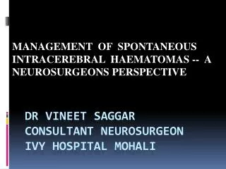
MANAGEMENT OF SPONTANEOUS INTRACEREBRAL HAEMATOMAS -- A NEUROSURGEONS PERSPECTIVE. DR vineet saggar Consultant neurosurgeon ivy hospital mohali. Spontaneous intracerebral hemorrhage (ICH) accounts for 9% to 25% of all strokes and has devastating consequences.

E N D
MANAGEMENT OF SPONTANEOUS INTRACEREBRAL HAEMATOMAS -- A NEUROSURGEONS PERSPECTIVE DR vineetsaggarConsultant neurosurgeonivy hospital mohali
Spontaneous intracerebralhemorrhage (ICH) accounts for 9% to 25% of all strokes and has devastating consequences. • More than 50% of patients die, and half of the survivors are left severely disabled. • Death at 1 year varies by different location: 51% for deep, 57% for lobar, 42% for cerebellar and 65% for brain stem hemorrhages Sudlow CL, etal. Stroke. 1997;28:491–499. Thrift AG, Dewey HM, MacdonellRA,etal.(NEMESIS). Stroke. 2001;32:1732–1738.
CAUSES OF SPONTANEOUS ICH • Depending on the underlying cause of hemorrhage, ICH may be classified as A- Primary when it originates from the spontaneous rupture of small arterioles damaged by chronic hypertension or cerebral amyloidangiopathy, representing at least 85% of all cases or B- secondary when associated with vascular malformations, • Bleeding related to an ischemic stroke, • tumors, • abnormal coagulation or vasculitis
Pathophysiology • Intracerebral haemorrhage due to chronic hypertension accounts for about one-half of the cases. • The underlying pathology is haemodynamic injury to perforating arteries, 100—400 urn in diameter, which arise directly from much larger trunks to enter the brain at right angles and are end arteries. • Whereas the cortical vessels are protected by a thicker smooth muscle layer in the media, a series of bifurcations and collateral vessels, the perforating arteries are subjected directly to changes in blood pressure.
The arteries in question include the lenticulostriate arteries, the thalamoperforating arteries the paramedian branches of the basilar artery and the superior and anterior inferior cerebellar arteries • The pathological lesions may take the form of hyalinosis, lipohyalinosis or focal necrosis and Charcot-Bouchard/miliary aneurysm formation
Locations of hypertensive ICH are as follows: • 65% in the basal ganglia, • 15% in the subcortical white matter, • 10% cerebellar • 10% pontine
MANAGEMENT • It is important to determine the underlying aetiology rapidly. A history of hypertension, drug abuse and anticoagulant treatment is important.
If a history of hypertension is not available, it may be difficult in the acute state in a patient with high blood pressure to decide whether it is due to previously undetected hypertension or secondary to raised intracranial pressure (ICP) with a Cushing response. • Signs of end organ damage (brain, retina, heart and kidneys) can help differentiate the two.
DIAGNOSTIC MODALITIES CT is the investigation of choice Advantages - CT scan evaluates the size and location of the hematoma, - Extension into ventricular system ,- Degree of surrounding edema, - Anatomical disruption (Class I, Level of Evidence A).
Hematoma volume may be easily calculated from CT scan images by use of the following formula ABC÷2 a derived formula from the calculation of the volume of the sphere.
Rt Basal Ganglia Haematoma With Intraventricular Extension in a Hypertensive
NEUROSURGEONS PERSPECTIVE • When do we need to be careful and evaluate further: • Subarachnoid hemorrhage, • IVH,(IntraventricularHaemorrage) - Underlying calcification, or - lobar hemorrhage without extention to basal ganglia. - Nonhypertensive younger patients
Case 1 • Young 22yr male presented with sudden onset severe headache followed by loss of consiousness. • Gcs at presentation was E1V1 M4, Pupils were equal reacting • Patient was immediately intubated and sent for CT Head
- By the time CT was completed and decision on angiography was made • Patients GCS had deteriorated to 5 and left pupil was dilated • Since CT angio at 3 am at night would have taken another 1 hour or so it was decided to operate patient without angiography for removal of clot and aneurysm clipping
CT ANGIOGRAPHY • CT-angiography is not routinely performed in most centers,but may prove helpful in predicting hematoma expansion and outcomes . • In a recent prospective study of 39 patients with spontaneous ICH, focal enhancing foci (contrast extravasation, ‘spot sign’) seen in initial CT-angiography were associated with the presence and extent of hematoma progression
Contrast extravasation seen in the hematoma of a patient with acute coagulopathicintracerebralhemorrhage (white arrows).
Conventional angiography • Required only in selected cases where diagnosis is in doubt or Ct angio is in conconclusive. • But still it remains the gold standard for diagnosis of non hypertensive and vascular causes of spontaneous ICH.
Once diagnosis is confirmed • Patients can be divided into two broad groups A. Spontaneous ICH with vascular pathology B. Spontaneous ICH without Vascular cause
Spontaneous ICH without vascular pathology Common causes are: • Hypertensive bleeds • Cerebral amyloidangiopathy • Coagulation disorders & use of anticoagulants ,thrombolytic agents.
Intracerebral haemorrhage due to chronic hypertension accounts for about one-half of the cases of NON VASCULAR CAUSES OF SPONTANEOUS ICH
Management in cases of hypertensive bleeds After establishment of diagnosis the next step is 1 . Airway control, breathing, and circulation Intubation is generally recommended if GCS of patient is less than or equal to 8 - Apart from that haemodynamic instability Is another indication.
2. Blood pressure control • Extreme levels of BP after ICH should be aggressively but carefully treated to reduce the risk of hematoma expansion and to keep and maintain cerebral perfusion pressure (CPP;CPP = mean arterial pressure (MAP) - ICP)
In general, the American Heart Association Guidelines indicate that systolic blood pressures exceeding 180 mmHg or MAP exceeding 130 mmHg should be managed with continuous infusion antihypertensive agents such as labetalol,esmolol, or nicardipine
The following algorithm adapted from guidelines from AHA may be helpful: 1. If systolic BP is >230 mm Hg or diastolic BP > 140 mm Hg on 2 readings 5 minutes apart, institute nitroprusside. 2. If systolic BP is 180 to 230 mm Hg, diastolic BP 105 to 140 mmHg, or mean arterial BP =130 mm Hg on 2 readings 20 minutes apart, institute intravenous labetalol, esmolol, enalapril, or other smaller doses of easily titratable intravenous medications such as diltiazem, lisinopril, or verapamil.
3. If systolic BP is ,<180 mm Hg and diastolic BP ,<105 mm Hg, defer antihypertensive therapy. Choice of medication depends on other medical contraindications (eg, avoid labetalol in patients with asthma). 4. If ICP monitoring is available, cerebral perfusion pressure should be kept at >70 mm Hg.
Low blood pressure • Volume replenishment is the first line of approach. Isotonic saline or colloids can be used and monitored with central venous pressure or pulmonary artery wedge pressure. If hypotension persists after correction of volume deficit, continuous infusions of pressors should be considered, particularly for low systolic blood pressure such as ,90 mm Hg. • Phenylephrine 2–10 mg / kg/ min • Dopamine 2–20 mg / kg/ min • Norepinephrine Titrate from 0.05–0.2 mg / kg / min
Management of Increased ICP ICP is considered a major contributor to mortality after ICH;thus, its control is essential. ICP may be managed through • Osmotherapy, • Controlled hyperventilation, • Barbiturate coma.
Elevated ICP is defined as intracranial pressure >20 mm Hg for .5 minutes. • A therapeutic goal is to achive ICP <,20 mm Hg and cerebral perfusion pressure (CPP) >70 mm Hg
Osmotherapy - The first medical line of defense is osmotherapy. However, it should not be used prophylactically. - Mannitol 20% (0.25– 0.5 g/kg every 4 h) is reserved for patients with • type B ICP waves, • progressively increasing ICP values, or • clinical deterioration associated with mass effect.
Due to its rebound phenomenon, mannitol is recommended for only 5 d. • To maintain an osmotic gradient, furosemide may be administered simultaneously • Serum osmolality should be measured twice daily and targeted to 310 mOsm/L.
Hyperventilation • Hypocarbia causes cerebral vasoconstriction. • Reduction of cerebral blood flow is almost immediate, • Although peak ICP reduction may take up to 30 minutes after pCO2 is changed. • .
Maintain pCO2 to 35–30 mm Hg, • best achieved by raising ventilation rate at constant tidal volume (12–14 mL/kg), lowers ICP 25% to 30% in most patients • . Failure of elevated ICP to respond to hyperventilation indicates a poor prognosis
Muscle relaxants • Neuromuscular paralysis in combination with adequate sedation can reduce elevated ICP by preventing increases in intrathoracic and venous pressure associated with coughing, straining, • Nondepolarizingagents,such as vecuronium or pancuronium, • Patients with critically elevated ICP should be pretreated with a bolus of a muscle relaxant before airway suctioning. • Alternatively, lidocaine may be used for this purpose.
NEUROSURGEONS PERSPECTIVE SURGERY When to operate when not to operate
A spectrum of ICH patients 1.Alert patients with subtle neurological signs and small (< 2 cm) haematomas Surgery not indicated 2.Indications for surgery between 1 and 3 are controversial The following patients are more likely to be operated upon (i) clot volumes between 20-80 ml,(2)superficial lobar haemorrhages, and (3) worsening conscious level/neurological deficit 3.Large haemorrhage with significant neuronal destruction and poor neurological status (GCS < 5)Surgery not indicated.
Most neurosurgeons would operate on patients with a deteriorating conscious level and a worsening neurological deficit • In addition, lobar/superficial haematomas 20—80 ml in volume are more likely to be operated upon • . Cerebellar haemorrhage greater than 3—4 cm should be operated upon, especially when there is concomitant clinical deterioration or hydrocephalus3
STICH I TRIAL • The need to gain robust evidence to support clinical decision making led to the initiation of the Surgical Trial in Intracerebral Haemorrhage (STICH) funded by the MRC and the Stroke Association in 1998.
STICH was a prospective randomised trial to compare Early surgery with Initial conservative treatment in with spontaneous supratentorialintracerebral haemorrhage.
A parallel group-trial design was used . Early surgery combined haematoma evacuation (within 24 hours of randomisation) with best medical treatment. Initial conservative treatment used best medical treatment although delayed evacuation was allowed if it became necessary. Analysis was on an intention-to-treat basis.
Primary Outcome Measure: 8 point Glasgow Outcome Scale sent as a postal questionnaire to patients at 6 months follow-up. From this a dichotomised prognosis-based outcome measure was used
Results: This trial was the largest to date and successfully recruited 1033 patients from 87 centres around the world. Patients were randomised to early surgery (503) or initial conservative treatment (530). Of 468 patients randomised to early surgery 26% had a favourable outcome compared to 24% of the 496 randomised to initial conservative treatment (95% CI 0.66-1.19, p=0.414). (Outcome data was unavailable for 69 patients).