B cell development
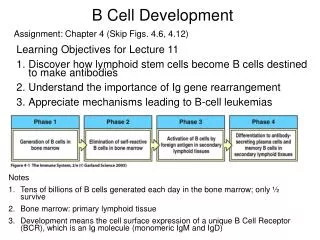
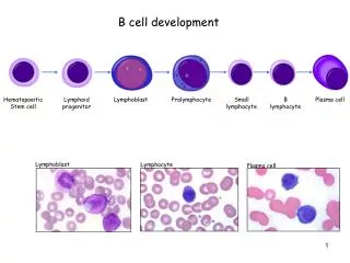
B cell development. Hematopoeitic Stem cell. Lymphoid progenitor. Lymphoblast. Prolymphocyte. Small lymphocyte. B lymphocyte. Plasma cell. Lymphoblast. Lymphocyte. Plasma cell. Clonal development of B cells. Clonal selection by antigen. Clonal proliferation (T cell help).

B cell development
E N D
Presentation Transcript
B cell development Hematopoeitic Stem cell Lymphoid progenitor Lymphoblast Prolymphocyte Small lymphocyte B lymphocyte Plasma cell Lymphoblast Lymphocyte Plasma cell
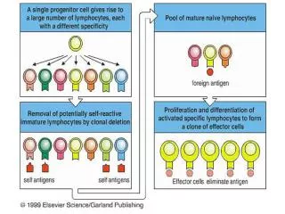
Clonal development of B cells Clonal selection by antigen Clonal proliferation (T cell help) Differentiation and maturation with T cell help plasma cells (antibody secretion) memory cells (recirculate)
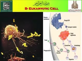
Plasma cells are antibody factories Lymphocyte Plasma cell golgi rough endoplasmic reticulum
Virgin B cells • Key facts • Short-lived cells (over 75% do not reach the circulation and die by apoptosis and are phagocytosed by bone marrow macrophages) • When they leave the bone marrow they are already antigen-specific • Have rearranged variable region genes • Express both IgM and IgD at the cell surface
The basic structure of immunoglobulin Variable domain Heavy chain Light chain V VH D V J Fab J VL CH1 CL CH2 Hinge region Fc CH3 Constant domain CH4 (some antibody classes only)
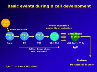
Variable region genes are rearranged in the bone marrow Lymphoid progenitor Immature B cell Pro-pre B cell Pre B cells Stem cell Cm+ Cm+ sIgM+ Heavy chain Vm -region gene rearrangement Light chain V-region (k or l) gene rearrangement Generation of antigenic specificity Variable region genes undergo rearrangement Independent of T-cell help Independent of antigen
The generation of antibody diversity VH V D V J J VL CH1 CL CH2 CH3 variable region constant region ~4 mheavy chain dheavy chain ~100 ~20 ...g1, g2, e, a CH1 J2 CH2 CH3 CH4 V3 J3 J4 V1 Vn V2 J1 CH1 CH2 CH3 CH4 D1 D2 D3 Dn V4 germline DNA mheavy chain dheavy chain ...g1, g2, e, a CH2 CH3 CH4 CH1 CH1 CH2 CH3 CH4 V2 D1 J3 rearranged DNA splice site primary mRNA transcript V2D1J3 m d spliced mRNA transcript d m V2D1J3 V2D1J3 mRNA for IgM mRNA for IgD
Class (isotype) switching rearranged DNA V2D1J3 m g1 g2 a d e mRNA for IgM, IgD class switch further DNA rearrangement V2D1J3 g1 class switch to IgG1 e g2 a mRNA for IgG1 class switch to IgE V2D1J3 a e mRNA for IgE A B cell that has undergone a class switch from IgM to IgG1 can still subsequently undergo a further class switch to IgE, IgA etc., however a B cell that has undergone a class switch to IgA cannot switch back to IgG1 or IgE, all heavy chain genes preceding the a chain gene ( g1, g2 and e) are excised during class switching.
Gene rearrangements during B cell development Variable region gene rearranged to generate “VDJ” (heavy chain) and “VJ” (light chain). Allelic exclusion prevents rearrangement of 2nd chromosome. IgM and IgD heavy chains produced - mature "virgin" B cell Isotype (class) switching - occurs following stimulation of virgin B cell with antigen Affinity maturation - further mutation of variable region, selection for high affinity antibodies.
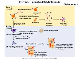
Events which follow encounter with antigen - the primary response Antibody formation (IgM) Virgin B cell antigen activation Expression of lymphocyte homing receptors, targets B cell to secondary lymphoid organs. Memory B cell pool quiescent The growth and proliferation phase requires "help" from T cells in the form of cytokines.
The secondary response Cells capable of antigen presentation include B cells, macrophages and follicular dendritic cells. Memory cells Antigen presenting cell (APC) Memory B cell CD4 Plasma cells producing IgG, A, E or M MHC class II TCR Helper T cell (Th -cell) The secondary response is very rapid and produces large quantities of high affinity antibody, predominantly IgG.
recovery threshold 4 days 2 weeks The primary and secondary antibody responses are different Primary exposure Secondary exposure to antigen to antigen Primary Secondary response response 100,000 IgG 10,000 1000 Log antibody titre 100 10 IgM 1 0 0 7 14 21 28 35 42 Days
Germinal centres Th cell B cell encounter with antigen secondary lymphoid follicle centrocyte plasma cell formation (IgM) follicular dendritic cell feline lymph node stained with a B cell-specific antibody centroblast Harris, N. L. ASH Image Bank 2002;2002:100368 plasma cells migration to: medullary chords of lymph nodes red pulp of spleen lamina propria of GALT lymphoblasts centroblast centrocyte class switching somatic hypermutation affinity maturation memory B cells continual re-circulation
B cell development in the secondary lymphoid tissues How long do B cells live? IgM+ IgD+ to lymphoid tissue (homing receptors) exits bone marrow germinal centre formation encounter with antigen primary lymphoid follicle mature B cell failure to access secondary lymphoid tissue no interaction with antigen - half-life of 3-8 weeks plasma cell formation (IgM) Cell death (life span of 2-3 days) IgM low IgD+ exits bone marrow to secondary lymphoid tissue retention in T cell area - APOPTOSIS Long-lived (years?) anergic B cell
What is B cell anergy? IMMATURE B CELL BONE MARROW no self- reaction sIgM+ sIgM+ sIgM+ soluble self-antigen multivalent self-antigen CLONAL DELETION sIgM+ sIgD+ sIgM+ low sIgD+ MATURE B CELL ANERGIC B CELL PERIPHERY
Improving the B cell response “Affinity Maturation” Th cell B cells encounter with antigen secondary lymphoid follicle centrocyte plasma cell formation (IgM) follicular dendritic cell centroblast affinity B cell with Ig of “highest affinity” Level antigen Follicular dendritic cell has antigen trapped on its surface Time post-immunisation/infection
Affinity is increased by “somatic hypermutation” mheavy chain dheavy chain ...g1, g2, e, a CH2 CH3 CH4 CH1 CH1 CH2 CH3 CH4 V2 D1 J3 rearranged DNA Somatic hypermutation* mheavy chain dheavy chain ...g1, g2, e, a CH2 CH3 CH4 CH1 CH1 CH2 CH3 CH4 V2 D1 J3 mutated DNA Higher affinity = Survival Lower affinity = Cell death
A short history of B cell development Bone marrow enters circulation Lymphoid tissue IgM Affinity maturation Class switching IgD virgin B cell (defined antigenic specificity) IgG – producing plasma cell Low affinity Low Avidity High titre IgM – producing plasma cell Low affinity High Avidity Low titre IgG – producing plasma cell High affinity Low Avidity High titre
Summary - Lymphocytes and their function(s) B1 cells Predominantly producing low affinity IgM. Immunity to polymeric antigens on microorganisms? Primitive immunity during foetal development? B2 cells Classical B cells producing all isotypes of immunoglobulins. High affinity immunoglobulins.
Bone marrow A Rough Guide to the humoral immune response Helper T cell cytokines No encounter with antigen = cell death antigen presentation to T cell e.g. interdigitating cell enters circulation IgM virus IgD IgM antigen presenting cell e.g.follicular dendritic cell in follicle Encounters antigen IgD virgin B cell (defined antigenic specificity) cytokines vaccine T / B cell cooperation B cell MHC class II IgG,E,A or M TCR high affinity antibody cytokines Helper T cell plasma cell booster IgM primary response then later IgG etc. migration or antigen presentation in lymphoid tissue plasma cell memory B cell memory B cell re-infection