Download
1 / 50
1.51k likes | 6.79k Vues
Disorders of Pigmentation. Skin colour. Determined by - melanin - haemoglobin - carotenoids Melanin - major determinant Melanin is synthesized by melanocytes within melanosomes and transferred to keratinocytes

E N D
Skin colour • Determined by - melanin - haemoglobin - carotenoids • Melanin - major determinant • Melanin is synthesized by melanocytes within melanosomes and transferred to keratinocytes • Constitutive skin colour - genetically determined • Facultative skin colour - induced by sun and hormones
Melanocyte • Dendritic cells • Derived from neutral crest • Migrate to epidermis • Epidermal melanin unit - one melanocyte connected to about 36 keratinocytes by dendrites • Synthesize melanin in organelles called melanosomes • Melanosomes are then transferred to keratinocytes through dendrites
Melanin • Two types - eumelanin (black or brown) - pheomelanin (reddish) • Derived from tyrosine Tyrosine Tyrosinase DOPA[3,4 dihydroxy phenylalanine] Dopaquinone Dopachrome 5, 6 dihydroxindole Eumelanin Cysteinyldopa pheomelanin
Disorders of pigmentation - An Overview • Skin pigmentation has far-reaching social and psychological implications • White people strive for tanning which while brown and black people strive for a lighter skin • Melanin pigmentation disorders are important for medical and cosmetic reasons
Classification • Hypomelanosis: reduced or absent pigment eg. Vitiligo, Pityriasis alba • Brown hypermelanosis: increased pigment in epidermis. eg. Freckles • Slate gray or blue hypermelanosis: pigment in dermis. eg. Mongolian spots
Approach to a patient: History • Onset : birth, infancy or later • Cause: sun exposure, drugs, occupation • Systemic complaints • Family history: neurofibromatosis, tuberous sclerosis, vitiligo
Approach to a patient: Examination: • Type of lesion: brown, blue, hypopigmented (check sensation), depigmented • Shape: Ash leaf macules (tuberous slerosis) Koebner phenomenon(vitiligo) • Distribution pattern : linear/segmental (nevus depigmentosus), symmetric (vitiligo), specific sites (melasma, Addison’s disease)
Approach to a patient: Examination aids: • Hand lens • Oblique lighting for elevation or depression • Dermatoscopy • Wood’s lamp - 360 nm. Epidermal pigmentary anomalies made more prominent • Histology- H and E for presence or absence of melanin Dopa reaction - melanocytes stain dark Silver stains - melanin stains black
Disorders of decreased pigmentation • Hypopigmentation • Depigmentation The pigment may be absent from birth or lost later in life. It may be diffuse or cicumscribed.
Classification: Hypogimented / depigmented lesions • Genetic and Developmental: Albinism, Nevus depigmentosus, Nevus anaemicus, Halo nevus, Tuberous sclerosis (ash leaf macule) • Endocrine: Addison’s disease, Hypothyroidism, Hypopituitarism • Nutritional: Vit.B12 deficiency, Kwashiorkor, Malabsorption
Classification: Hypogimented / depigmented lesions • Post-inflammatory: Pityriasis alba, Eczema, Psoriasis, Pityriasisrosea, Lupus erythematosus, Morphea, Scleroderma, Bullousdermatoses • Infection: Leprosy, Tineaversicolor, Candidiasis, Post kalaazar dermal leishmaniasis • Chemicals and Drugs: Phenols, Arsenicals, Hydroquinone, Steroids
Classification: Hypogimented / depigmented lesions • Physical: Burns, Trauma, Post dermabrasion, Post laser • Miscellaneous: Idiopathic guttatehypomelanosis, Vitiligo, Mycosis fungoides
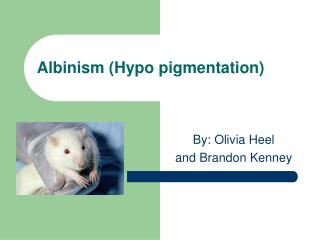
Albinism • Oculocutaneous albinism involves skin, hair and eyes • Mostly autosomal recessive • Absence of pigmentation from birth • Photophobia, reduced visual activity • Sunburns, skin cancers common • Protection of eyes and skin by sunglasses, sunscreens SPF > 20, clothing
Waardenburg’s syndrome • Rare, autosomal dominant disorder • White forelock • Hypertelorism • Congenital deafness • Hypomelanoticmacules • Heterochromicirides • Incomplete forms may occur
Tuberous sclerosis • Autosominal dominant, neurocutaneous syndrome with skin lesions, mental retardation and epilepsy • Skin lesions are ash-leaf macules, angiofibromas and shagreen patches • Ash-leaf macules - present at birth in>90% cases, so important in early diagnosis Oval or ash-leaf shaped, hypopigmentedmacules, made prominent in Wood’s lamp • Long axis is axial on limbs and transverse on trunk
Nevus depigmentosus • A hypopigmented birthmark which is congenital and stable • Irregular, geographic margins and quasidermatomal distribution • Block in transfer of melanosomes from melanocytes to keratinocytes • Sporadic occurrence, no medical significance and no treatment required
Kwashiorkor • Protein deficiency in post weaning years • Reddish patches which turn into dark plaques which turn white after exfoliation (crazy pavement dermatosis) • Disruption of melanogenesis is due to multiple deficiencies • Pigment changes and dyschromic hair are reversible with proper diet
Tineaversicolor • Common, superficial fungal infection • Overgrowth of Malasezziafurfur - a normal resident • Common after puberty; face, neck, upper trunk affected • Nonpruritic or mildly pruritic, hypo or hyperpigmented lesions with fine scales • Common in tropics; during summers
Leprosy • Both hypopigmented and erythematous lesions common • Hypopigmentedmacules common in tuberculoid type of disease • Each hypopigmented lesions in leprosy endemic areas should be examined for sensations of touch, pain, temperature • Treatment according to type of leprosy
Pityriasis alba • A common disorder in children • Hypopigmented lesions with powdery scaling; chiefly affecting face • Etiology not known but may be a feature of atopy or malnutrition • To be differentiated from indeterminate leprosy and early vitiligo • Treatment with emollients
Other causes • Chemical, thermal burns, trauma • Postinflammatory: after healing of lesions of eczema, psoriasis, discoid lupus erythematosus, lichen striatus etc. • Endocrine: hypopitutarism and hypogonadism - diffuse hypomelanosis • Idiopathic guttatehypomelanosis: tiny depigmentedmacules an extremities due to ageing
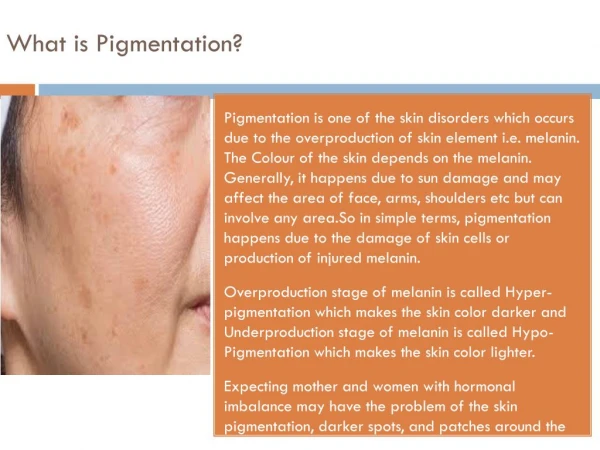
Disorders of hyperpigmentation • May be epidermal or dermal • Epidermal hyperpigmentation due to - Increased melanin with normal number of melanocytes - Increased number of melanocytes • Dermal hyperpigmentation due to - Melanin from epidermis transferred to dermis - Melanin formed in dermal melanocytes - Melanin pigments appears blue-gray due to Tyndall effect
Classification: Epidermal hyperpigmentation • Physiologic: Pigmentary demarcation lines, suntanning • Genetic and Developmental: Lentigines, Freckles, Peutz-Jeghers syndrome, Melanocytic nevus, Café-au-lait spots, Xerodermapigmentosum, Becker’s nevus, Nevus spilus, Acanthosisnigricans
Classification: Epidermal hyperpigmentation • Post-inflammatory: Eczema, Psoriasis, Lichen planus, Lupus erythematosus, Scleroderma, Morphoea, Vagabond’s disease • Infection: Tineaversicolor, Tineanigra • Nutritional: Kwashiorkor, Pellagra, Vit.B12, Vit.C, Folic acid deficiency
Classification: Epidermal hyperpigmentation • Physical: Trauma, Radiation dermatitis • Endocrine: Melasma, Addison’s disease, Cushing’s syndrome, Phaeochromocytoma, Acromegaly, Hyperthyroidism • Neoplastic: Malignant melanoma, Seborrhoeickeratosis, Pigmented basal cell carcinoma
Classification: Dermal hyperpigmentation • Genetic and Developmental: Mongolian spots, Nevus of Ota/Ito, Incontinentiapigmenti • Inflammatory: Stasis dermatitis, Post inflammatory to eczema and fixed drug eruption • Chemicals and Drugs: Anti-malarials, OC Pills, Minocycline, Clofazimine, Topical hydroquinone, Tattoos
Classification: Dermal hyperpigmentation • Endocrine: Melasma • Physical: Thermal burns, Post traumatic • Infection: Syphilis, Yaws, Pinta • Neoplastic: Metastasis of melanoma
Classification: Dermal hyperpigmentation • Nutritional: Chronic nutritional deficiency • Metabolic: Hemochromatosis, Alkaptonuria, Macular/Lichen amyloidosis • Miscellaneous: Pigmented purpuricdermatosis, Purpura
Melanocytic nevi • Benign proliferations of melanocytic nevus cells at the dermo-epidermal junction • May be congenital or acquired • Acquired nevi are more common • Appear in infancy or childhood, slowly grow and mature and then regress in older life • Important for cosmetic reasons and as precursors for melanoma (esp in white)
Acquired nevi • Round or oval, uniformly coloured and sharply bordered lesions • Appear after birth • Increase in frequency during childhood and adolescence and plateaus during middle age • Most of them start as junctional nevi which are flat and histologically confined to dermal-epidermal junction
Acquired nevi • Gradually mature to compound nevi which have nests and columns of nevus cells in dermis along with the junctional component. These are raised, rounded, brown or black • Intradermal nevi : Compound nevi mature to intradermal nevi with nevus cells only in dermis having neuron like appearance. These are dome shaped, nonpigmented and may have one or more coarse hairs
Treatment Elliptical excision and biopsy • Indications of excision: • Cosmetic reasons • Irritation due to clothing, belts, straps • Atypical appearance: sudden increase in size, varied pigmentation, irregular borders, bleeding. Destructive methods (cautery, cryotherapy) not recommended as recurrence with atypical appearance may occur
Congenital melanocytic nevi • Small < 1.5 • Intermediate: 1.5 to 20 cms • Giant > 20 cms • Malignant potential for giant nevi is 4-6% • Excision justified for cosmetic reasons and risk of malignancy
Café au lait macules (CALM) • Circumscribed, brown macules with irregular margins, 2-5 cm in size • Present at birth • Isolated CALM may occur in 10-20% of normal population • No increase in the number of melanocytes • Five or more CALM of size >0.5 cm in prepubertal age group and >1.5 cm in an adult are strongly suggestive of neurofibromatosis
Becker’snevus • Acquired, pigmented, hairy plaque common on trunk, more common in males • Appears in first or second decade • Common sites: shoulder, chest, back • May become verrucous with hair growth and then remains stable • No treatment needed
Ephelides (Freckles) • Tiny (<0.5 cm), discrete brown macules • Common in fair skinned • Appear in childhood on sun exposed parts; lighten in absence of sun exposure • Melanocytes are not increased in number but are hyperactive • May be part of some syndromes
Melasma • A common macular brown coloured lesion seen on face in males and females • Common in pregnancy: Mask of pregnancy • Etiology: hormones (OC Pills) and sunlight • May disappear or remain after delivery • Forehead, nose, cheeks affected • Exacerbation on sun exposure • Histologically may be epidermal, dermal or mixed
Treatment of melasma • Sunscreens and bleaching agents • Physical sunscreens to be used daily • Bleaching agents : Hydroquinone 2-4%, Kojic acid 2-3%, Tretinoin 0.025-0.05%, Arbutin • About two months needed to see a response and six months for complete resolution • Recurrence common after sun exposure • Dermal melasma does not respond
Diffuse hyperpigmentation • Addison’s disease • Haemochromatosis • HIV infection and AIDS • Drugs: Clofazimine Chlorpromazine Amiodarone Anticancer agents
Post-inflammatory hyperpigmentation • After resolution of specific eruptions • Common after lichen planus, atopic dermatitis, acne vulgaris, contact dermatitis, psoriasis, pyodermas etc. • Discrete macules exactly on the sites previously affected by eruptions • May persist for months
Fixed Drug Eruption (FDE) • NSAIDs, antibiotics, barbiturates etc. • Reddish brown macule → edematous → desquamation → pigmentation • Recurs at same site on rechallenge • May become generalised or blistering • Melanin is increased in epidermis and dermis (melanophages)
Mongolian spots • Common in asian newborns on buttocks or lower back • Due to arrest of migrating melanocytes in the dermis • No treatment is needed as they spontaneously disappear by 2 to 10 yrs of age
Nevus of Ota and Ito • Nevus of Ota (around eyes) and nevus of Ito (shoulder area) are other examples of dermal pigmentation
Drugs Hydroquinone (2 - 4%) • Inhibits tyrosinase activity by 90% • Affects DNA, RNA synthesis • Safety concerns like irritation, exogenous ochronosis • May be combined with topical steroids, tretinoin, kojic acid Tretinoin • Adjuvant • Inhibits tyrosinase in cells cultures • Exact mechanism of action unknown
Drugs Kojic acid • Fungal metabolite • Food additive - Japan • Suppresses tyrosinase by chelating copper • Stability • Sensitization A.H.A • Natural saturated dicarboxylic acid • Diminished corneocyte cohesion • Faster desquamation due to increased turnover • Side effects: irritation, hyperpigmentation • Glycolic, lactic, mandelic acid
Drugs Azelaic acid • Antiproliferative, cytotoxic • Inhibits tyrosinase • As effective as hydroquinone • Azelaic acid 20% + Glycolic acid 15-20% is an effective combination Vit. C Problems with penetration and stability
Physical modalities • Chemical peels with increasing concentrations of glycolic acid, trichloroacetic acid, salicylic acid • Lasers : • Q switched Nd: YAG laser • Alexandrite laser • Pulse dye laser • Intense pulse dye laser